 Taste
TasteIN THIS CHAPTER:
 Taste
Taste
 Smell
Smell
 Structure and Function of the Eye
 Structure and Function of the Ear
 Solved Problems
Sensory organs are specialized extensions of the nervous system that contain sensory (afferent) neurons adapted to respond to specific stimuli and conduct nerve impulses to the brain. Sensory organs are very specific as to the stimuli to which they respond.
The senses of the body are classified as general senses or special senses. General senses include the cutaneous receptors (touch, pressure, heat, cold, and pain) within the skin that provide the sense of touch. Special senses are localized in complex receptor organs and have extensive neural pathways. The special senses are the senses of taste, smell, sight, hearing, and balance.
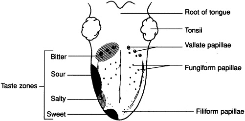
Receptors for the sense of taste (gustation) are located in taste buds on the surface of the tongue. The taste buds are associated with peglike projections of the tongue called lingual papillae (Figure 12-1). A few taste buds are also located in the mucous membranes of the palate and pharynx. A taste bud contains a cluster of 40 to 60 gustatory cells, each innervated by a sensory neuron, as well as many more supporting cells. The four primary taste sensation are sweet (evoked by sugars, glycols, and aldehydes); sour (evoked by H+, which is why all acids taste sour); bitter (evoked by alkaloids); and salty (evoked by anions of ionizable salts). Sensory innervation of the tongue and pharynx is by a branch of the facial nerve, CN VI, from the anterior 2/3 of the tongue, the glossopharyngeal nerve, CN IX, from the posterior 1/3 of the tongue, and the vagus nerve, CN X, from the pharyngeal region. Taste sensations are transmitted to the brain stem, then to the thalamus, and finally to the cerebral cortex, where taste perception occurs.


Figure 12-1. The surface of the tongue.
Receptors for the sense of smell (olfaction) are located in the nasal mucosa of the superior nasal concha. Like taste receptors, smell receptors are chemoreceptors, specialized neurons that respond to chemical stimuli and require a moist environment to function. The airborne chemicals become dissolved in the mucous layer lining the superolateral part of the nasal cavity. The olfactory nerve, CN I, transmits most impulses related to smell. Olfactory sensations are conveyed along each olfactory tract to the olfactory portions of the cerebral cortex where olfactory perception occurs.
Accessory structures of the eye either protect the eye or enable eye movement. These structures include the bony orbit, the eyebrow, the eyelids, the lacrimal apparatus (lacrimal glands that produce lacrimal fluid or tears, and the lacrimal canals and lacrimal sac, which drain the fluid into the nasal cavity), and the eye muscles (responsible for eye movements).
Muscles Involved in Eye Movements
Superior rectus rotates the eye superiorly
Medial rectus rotates the eye medially
Lateral rectus rotates the eye laterally
Inferior rectus rotates the eye inferiorly
Superior oblique rotates the eye inferolaterally
Inferior oblique rotates the eye superolaterally

The spherical eye is approximately 25 mm (1in.) in diameter. It consists of three tunics (layers), a lens, and two principal cavities (Figure 12-2).
Figure 12-2. The internal anatomy of the eye.
The fibrous tunic has two parts. The sclera is composed of dense regular connective tissue that supports and protects the eye and is the attachment site for the extrinsic eye muscles. The transparent cornea forms the anterior surface of the eye. Its convex shape refracts incoming light rays.
The vascular tunic has three parts. The choroid is a thin, highly vascular layer that supplies nutrients and oxygen to the eye and absorbs light, preventing it from being reflected. The ciliary body is the thickened anterior portion of the vascular tunic. It contains smooth muscle fibers that regulate the shape of the lens. The iris forms the most anterior portion of the vascular tunic and consists of pigment (that gives the eye color) and smooth muscle fibers arranged in a circular and radial pattern that regulate the diameter of the pupil, which is the opening in the center of the iris.
The receptor component of the eye contains two types of photoreceptors. Cones (approximately 7 million cones per eye) function at high light intensities and are responsible for daytime color vision and acuity (sharpness). Rods (approximately 100 million per eye) function at low light intensities and are responsible for night (black-and-white) vision. The retina also contains bipolar cells, which synapse with the rods and cones, and ganglion cells, which synapse with the bipolar cells. The axons of the ganglion cells course along the retina to the optic disc and form the optic nerve (CN II). The fovea centralis is a shallow pit at the back of the retina that contains only cones. It is the area of keenest vision. Surrounding the fovea centralis is the macula lutea, which also has an abundance of cones.
The lens is a transparent, biconvex structure composed of tightly arranged proteins. It is enclosed in a lens capsule and held in place by the suspensory ligament that attaches to the ciliary body. The lens focuses light rays for near and far vision.
The interior of the eye is separated by the lens into an anterior cavity and a posterior cavity (vitreous chamber). The anterior cavity is partially subdivided by the iris into an anterior and a posterior chamber. The anterior cavity contains a watery fluid called aqueous humor. The posterior cavity contains a transparent jellylike substance called vitreous humor.
 Warning!
Do NOT confuse the anterior and posterior chambers with the anterior and posterior cavity! The chambers are subdivisions of the anterior cavity.
The field of vision is what a person visually perceives. There are three visual fields, the macular field, the area of keenest vision, the binocular field, the portion viewed by both eyes, but not keenly focused on, and the monocular field, that area viewed by one eye and not shared by the other.
The neural pathway for vision consists of the light rays striking the photoreceptors in the retina, which causes the transmission of nerve impulses along the optic nerve to the optic chiasma. The optic tract, a continuation of optic nerve fibers from the optic chiasma, carries the impulses to the occipital cerebral lobes where vision occurs.
For an image to be focused on the retina, the more distant the object, the flatter must be the lens. Adjustments in lens shape, accomplished by the ciliary muscles in the ciliary body, are called accommodation. When these smooth muscles contract, the fibers within the suspensory ligament slacken, causing the lens to thicken and become more convex.
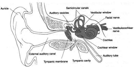
The ear is the organ of hearing and equilibrium. It consists of three principal regions: the outer ear, the middle ear, and the inner ear (Figure 12-3). The outer ear is open to the external environment and directs sound waves to the middle ear. Structures of the outer ear include the auricle (pinna), the external auditory canal, and the tympanic membrane (“eardrum”). The auricle directs the sound waves to the external auditory canal, a 2.5 cm (1 in.) fleshy tube that fits into the bony external acoustic meatus. The thin tympanic membrane conducts sound waves to the middle ear

The middle-ear cavity or tympanic cavity is the air-filled space medial to the tympanic membrane. The structures of the middle ear are the auditory ossicles, the auditory muscles, and the auditory (eustachian) tube. The auditory ossicles are three small bones that extend from the tympanic membrane to the vestibular (oval) window: the malleus (“hammer”); the incus (“anvil”); and the stapes (“stirrup”). These small bones amplify the sound waves. The auditory muscles are two tiny skeletal muscles that function reflexively to reduce the pressure of loud sounds before it can injure the inner ear. The auditory (eustachian) tube connects the middle-ear cavity to the pharynx. It functions to drain moisture from the middle ear cavity and to equalize air pressure on both sides of the tympanic membrane.
The inner ear contains the organs of hearing (cochlea) and equilibrium and balance (vestibular apparatus). The structures of the inner ear are described below. The bony labyrinth is a network of cavities that consist of three bony semicircular canals, the ampulla at the base of each semicircular canal, a central vestibule, and the cochlea. The membranous labyrinth is an intercommunicating system of membranous ducts seated in the bony labyrinth. Its parts are co-named with those of the bony labyrinth. The membranous semicircular canals and their ampullae posses receptors sensitive to rotary motions of the head. The vestibule consists of a connecting utricle and saccule, which possess receptors sensitive to gravity and linear motions of the head. These structures make up the vestibular apparatus. The membranous labyrinth is filled with a fluid, endolymph, and to the outside of the membranous labyrinth is a fluid called perilymph. The vestibular (oval) window, a membrane-covered opening from the middle ear into the inner ear, is located at the footplate of the stapes where it transfers sound waves from the solid medium of the auditory ossicles to the fluid medium of the cochlea. Within the cochlea is the membranous cochlear duct and the spiral organ (organ of Corti), the organs of hearing. The cochlear window (round window) is directly below the vestibular window that reverberates in response to loud sounds.

1. Sound waves are funneled by the auricle into the external auditory meatus.
2. The sound waves strike the tympanic membrane, causing it to vibrate.
3. Vibrations of the tympanic membrane are amplified as they pass through the malleus, incus, and stapes.
4. The vestibular window is pushed back and forth by the stapes setting up pressure waves in the perilymph of the cochlea.
5. The pressure waves are propagated to the endolymph contained within the cochlear duct.
6. Stimulation of the hair cells within the spiral organ of the cochlea causes the generation of nerve impulses in the cochlear nerve (CN VIII), which pass to the pons of the brain.
True or False
___1. Taste buds occur on the surface of the tongue, but are also found in smaller number in the mucosa of the palate and pharynx. (True)
___2. Contraction of the lateral rectus muscle rotates the eye laterally, away from the midline. (True)
___3. The anterior chamber is located between the cornea and the iris and is filled with vitreous humor. (True)
___4. The malleus is the bone in the middle ear that is attached to the vestibular window. (False)
___5. Foramina within the cribriform plate are associated with olfaction. (True)
Matching