 Lymphatic Structures
IN THIS CHAPTER:
 Lymphatic Structures
 Nonspecific Defenses
 Antibody-Mediated Immunity
 Cell-Mediated Immunity
 Transfusion Rejection Reaction
 Solved Problems
Functioning together with the cardiovascular system, the lymphatic system:
1. transports interstitial fluid, called lymph, in lymph vessels from the tissues back to the blood where it contributes to blood plasma;
2. assists in fat absorption in the small intestine; and
3. plays a key role in protecting the body from bacterial invasion via the blood.
Interstitial fluid enters the lymphatic system through the walls of lymph capillaries, composed of simple squamous epithelium. Lymph is carried then into larger lymphatic ducts. The lymph ducts eventually empty into one of two principal vessels: the right lymphatic duct, which drains lymph from the upper right quadrant of the body, and the large thoracic duct, which drains lymph from the remainder of the body. These drain into the right and left subclavian vein, respectively. Lymph flows through lymphatic vessels through the contraction of skeletal muscle, intestinal peristalsis, and gravity. Valves in lymphatic vessels prevent backflow.

Lymph nodes are small oval bodies enclosed in fibrous capsules. They contain phagocytic cortical tissue adapted to filter lymph. Afferent lymphatic vessels carry lymph into the node and efferent lymphatic vessels carry the filtered lymph from the node. Lymphocytes, leukocytes responsible for body immunity, and macrophages, phagocytic cells, are found in lymph nodes. Lymph nodes occur in clusters or chains. Some principal groups are:
• Popliteal and inguinal nodes of the lower extremity
• Lumbar nodes of the pelvic region
• Cubital and axillary nodes of the upper extremity
• Cervical nodes of the neck
• Mesenteric nodes associated with the small intestine
Lymphoid organs are:
• Tonsils: The three tonsils, (pharyngeal (adenoid), palatine, and lingual), are lymph organs of the pharyngeal region. They function to fight infections of the nose, ear, and throat region.
• Spleen: The spleen is located in the upper left portion of the abdominal cavity. It is not a vital organ in the adult but assists other organs in producing lymphocytes, filtering blood and destroying old erythrocytes. It also serves as a reservoir for erythrocytes.
• Thymus: The thymus is in the anterior thorax, deep to the manubrium of the sternum. It is much larger in a child than an adult. In a child it functions to change undifferentiated lymphocytes into T lymphocytes and is a reservoir for lymphocytes.
Types of non-specific defense mechanisms include:
• Mechanical barriers: skin and mucous membranes
• Chemical barriers: enzymes, HCl in stomach, lysozymes
• Interferon: proteins that inhibit viral growth
• Phagocytes: neutrophils, monocytes, and macrophages
• Species resistance
Specific immunity refers to resistance of the body to specific foreign agents (antigens). These include microorganisms, viruses, and their toxins, as well as foreign tissue and other substances. Antigens are large complex molecules (proteins, polysaccharides) found on cell walls or membranes of foreign substances.
In antibody-mediated immunity, an antigen stimulates the body to produce special proteins, antibodies, that destroy the particular antigen through an antigen-antibody reaction. Antibodies are gamma globulins composed of four interlinked polypeptide chains, two short (light) chains and two long (heavy) chains (Figure 17-1). All antibodies have constant regions, that are structurally similar, and variable regions, that are the location of antigen-binding sites. Small variations in the variable region make each antibody highly specific for one particular antigen. Antigen-antibody binding induces the production of more antibodies specific for that antigen.
Figure 1-1. A simple model of an antibody. Shaded areas indicate variable regions.
You Need to Know 
Active Immunity: The body manufactures antibodies in response to direct contact with an antigen. When that antigen is encountered again, the body “remembers” it and responds more quickly. This is how vaccinations work.
Passive Immunity: Result of the transfer of antibodies from one person to another, as a result of an injection or transfer across the placenta.
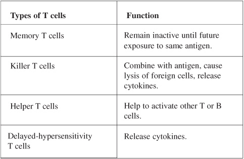
Cell-mediated immunity is another mechanism of specific immunity. In this case, cells provide the main defensive strategy. Lymphocytes, circulating in the blood and found in lymphoid tissues, (T lymphocytes and B lymphocytes) become sensitized to an antigen, attach themselves to that antigen, and destroy it. T lymphocytes produce cell-mediated immunity. Upon interacting with a specific antigen they become sensitized and differentiated in several types of daughter cells.
Table 17.1 Types and Functions of T Cells

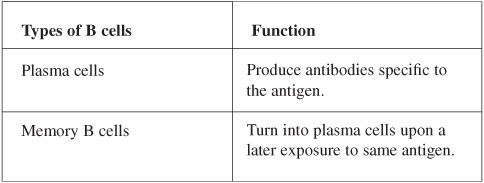
B lymphocytes produce antibody-mediated immunity. B lymphocytes become sensitized to an antigen, proliferate, and differentiate to form clones of daughter cells.
Table 17.2 Types and Functions of B Cells

Other components of the immune system are:
• Cytokines (interferons, chemotactic factors, macrophage-activating factors, migration-inhibiting factors, transfer factors): Chemical messengers used by the immune system to enhance immune response.
• Complement system: Enzyme precursors that aid immune response by causing lysis of invading cells, attract and enhance the action of phagocytes, enhance inflammation, and neutralize viruses.
Red blood cells have large numbers of antigens present on their cell membranes; these can initiate antibody production, and therefore antigen-antibody reactions. One of the groups of antigens most likely to cause blood transfusion reactions is the ABO system. Antigens are inherited factors present on the RBC membranes at birth. If the recipient of a blood transfusion and the donor are improperly matched the antigen-antibody reaction (transfusion reaction) occurs (see Table 17.3) causing RBC to clump, rupture, and hinder the flow of blood.
Table 17.3 The ABO Antigen System and Potential Donors

Another group of antigens associated with blood is the Rh system. Rh antigens are present on the red blood cell membranes of about 85% of the population. These people are classed as Rh positive (Rh+). The remaining 15% are classed as Rh negative (Rh-). Rh-individuals do not develop antibodies against Rh antigens until they are exposed to Rh+ blood.
True or False
____ 1. A person with type B blood has B antibodies. (False)
____ 2. A person who encounters a pathogen and who has a primary immune response develops passive immunity. (False)
____ 3. The interaction of antigen with antibody is highly specific. (True)
____ 4. Valves are present in lymphatic vessels. (True)
____ 5. Passive immunity is the transfer of antibodies developed in one individual into the body of another. (True)
____ 6. When antigenically stimulated, B lymphocytes proliferate and form plasma cells. (True)
____ 7. Antigens are small lipid molecules that stimulate the immune response. (False)