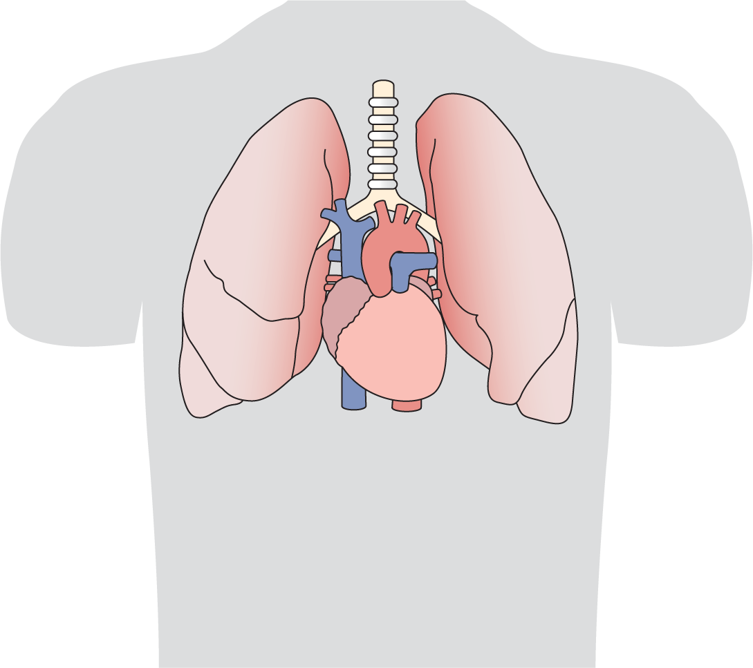
Figure 1-1 Location of the heart in the chest cavity.
© Jones & Bartlett Learning.
Since you are reading a book on electrocardiography, we assume that you have some basic knowledge of anatomy. However, a review is never a bad thing, so we are going to cover the basic anatomy of the heart and then concentrate on the electrical conduction system.
The heart sits in the middle of the chest at a slight angle pointing downward, to the left, and slightly anterior. Take a look at Figure 1-1.

Figure 1-1 Location of the heart in the chest cavity.
© Jones & Bartlett Learning.
Now, let’s look at the heart itself. First, from an anterior view, and then in cross section.
The right ventricle (RV) dominates the anterior view (Figure 1-2). Most of the anterior surface of the ventricles consists of the RV surface. A key point to remember is that, though the RV dominates this visual view, the left ventricle (LV) dominates the electrical view. We will review this in more detail in Chapter 4, Vectors and the Basic Beat, when we discuss vectors.

Figure 1-2 Anterior view of the heart.
© Jones & Bartlett Learning.
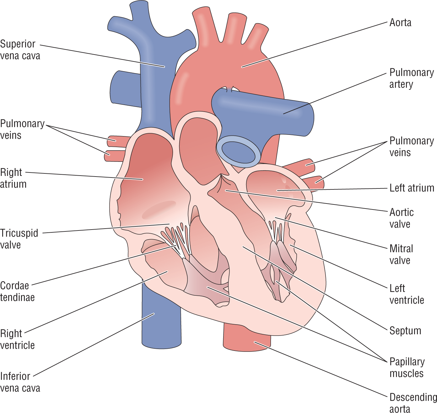
DescriptionFigure 1-3 shows a cross-sectional view of the heart. In the following sections, we will cover the function of the heart as a pump and review the electrical conduction system in greater detail.

Figure 1-3 Cross-sectional view of the heart.
© Jones & Bartlett Learning.
Description