Figure 4-6 Leads view the heart from different angles.
© Jones & Bartlett Learning.
Leads Are Like Pictures of the Heart
So, the electrodes (leads) pick up the electrical activity of the vectors, and the ECG machine converts them to waves. Think of each set of waves as a picture. Now, imagine that we place various electrodes—or cameras, for this analogy—at certain angles to the main axis (Figure 4-6). We would get multiple pictures of the heart from a three-dimensional perspective. Think of an ECG as a picture album, and it will be easy. To make matters more interesting, suppose we gave you multiple photographs of a toy elephant, including some reference for scale. Would you be able to put them together three-dimensionally in your mind’s eye? Of course you would! This is all an ECG is meant to give us a three-dimensional picture of the heart’s electrical axis. From this picture, we can get all sorts of information about where pathologic processes––such as infarcts, hypertrophy, and blocks––are occurring.

Figure 4-6 Leads view the heart from different angles.
© Jones & Bartlett Learning.
Lead Placement (Where to Put the “Cameras”)
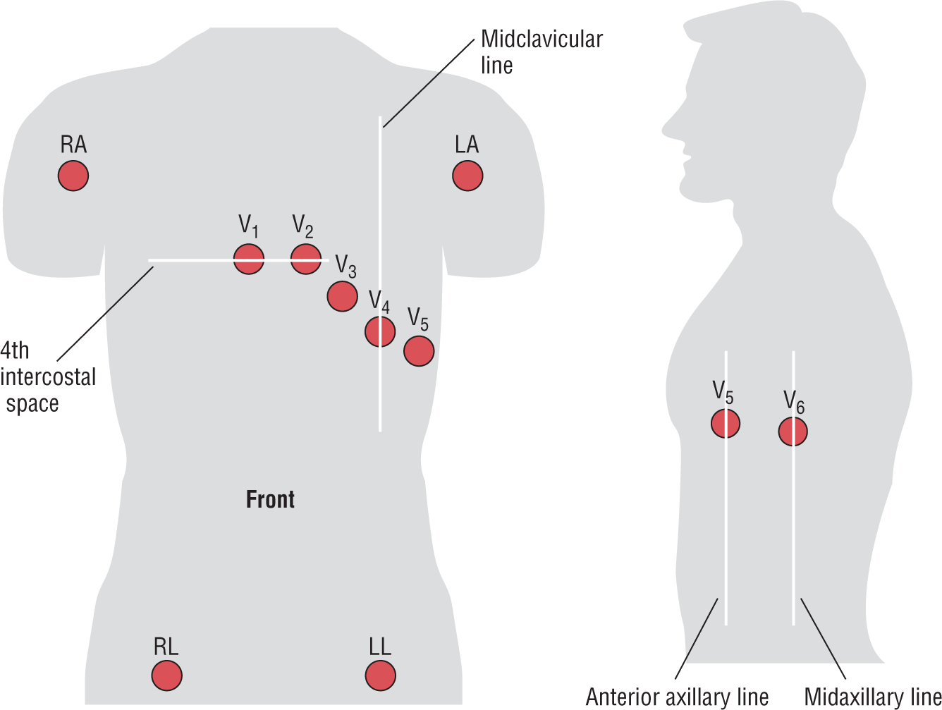
All right, so where do you place the cameras, or electrodes? You place them over the areas shown in Figure 4-7. The limb leads (extremity leads)—the right arm (RA), left arm (LA), right leg (RL), and left leg (LL)—are placed at least 10 cm from the heart. It doesn’t matter if you place the arm leads on the shoulders or the arms, as long as they are 10 cm from the heart. The precordial leads (chest leads), however, have to be placed exactly. Position V1 and V2 on each side of the sternum at the fourth intercostal space. To find the space, first isolate the Angle of Louis. This is a hump located near the top third of the sternum. Start feeling down your sternum from the top, and you’ll feel it. It is located next to the second rib. The space directly beneath it is the second intercostal space. Count down two more spaces and you’re there. V4 is at the fifth intercostal space in the midclavicular line. Follow the diagram for the remaining positions.

Figure 4-7 Lead placement.
© Jones & Bartlett Learning.
DescriptionHow the Machine Manipulates the Leads
The ECG machine reads the positive and negative poles of the limb electrodes to produce leads I, II, and III on the ECG (Figure 4-8). In other words, the camera is placed at the positive pole and aimed down the lead in question. In physics, two vectors (or in this case leads) are equal as long as they are parallel and of the same intensity and polarity. Therefore, we can move the leads from the locations shown in Figure 4-8 to a point passing through the center of the heart, and they will be the same (Figure 4-9A). By doing some complicated vector manipulation, the machine comes up with three additional leads (Figure 4-9B).

Figure 4-8 Leads I, II, and III.
© Jones & Bartlett Learning.
Description
Figure 4-9 Vector manipulation results in three more leads.
© Jones & Bartlett Learning.
Description