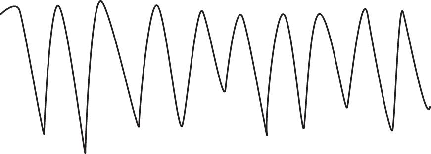
Figure 6-1 Artifact caused by a moving lead wire can result in a misdiagnosis of the rhythm.
© Jones & Bartlett Learning.
Artifacts are false abnormalities of the baseline present on an ECG or rhythm strip that are due to sources other than the patient’s bioelectrical impulses (Figure 6-1). The sources of these abnormalities can be anything from movement on the part of the patient, movement of the electrical leads (wires), muscle tremors, and interference from external electrical equipment.

Figure 6-1 Artifact caused by a moving lead wire can result in a misdiagnosis of the rhythm.
© Jones & Bartlett Learning.
Many times the artifact can be easily identified and discounted. Other times, the artifact can be mistaken for the patient’s rhythm or an arrhythmic event. In those cases, the artifact may lead to a misdiagnosis of the rhythm and result in inappropriate, sometimes dangerous, or unnecessary treatment.
In some circumstances, as in Figure 6-2, the entire rhythm strip can be altered by the artifact. In this example, the clinicians could easily have misdiagnosed the rhythm for a very dangerous rhythm known as ventricular tachycardia. Sometimes the artifact presents for only a short time, as in Figure 6-3. A quick way to verify your rhythm abnormality is to change the lead that your monitor is viewing, since many times the artifact will present in only one lead. If any questions remain, remember to view the “company that the arrhythmia keeps” (Figure 6-4). What we mean by this statement is that the arrhythmia does not occur in a void. Take a look at your patient and their clinical scenario before deciding on a course of action.

Figure 6-2 This patient was wrestling with her child. The movement of the leads caused the clinician to believe that the patient was in ventricular tachycardia. The rhythm returned to normal when the patient stopped moving.
© Jones & Bartlett Learning.

Figure 6-3 Interference by an electrical appliance in the room was the cause of this artifact. Turning off the machine caused the baseline to return to normal.
© Jones & Bartlett Learning.

Figure 6-4 No, this patient was not in an asystolic cardiac arrest! The lead fell off his chest. When in doubt, take a look at your patient. A person cannot be sitting up eating dinner and be in asystole (lack of any electrical activity in the heart) at the same time. Remember to always look at “the company that the arrhythmia keeps.”
© Jones & Bartlett Learning.
There is no easy way to tell what is artifact and what is the normal morphologic appearance of a rhythm. Unfortunately, it takes a lot of time and experience in strip interpretation before you get the hang of it. A word of wisdom is to always look at what is abnormal and concentrate on that spot. Usually, that is where the answer lies, even if it is just artifact.