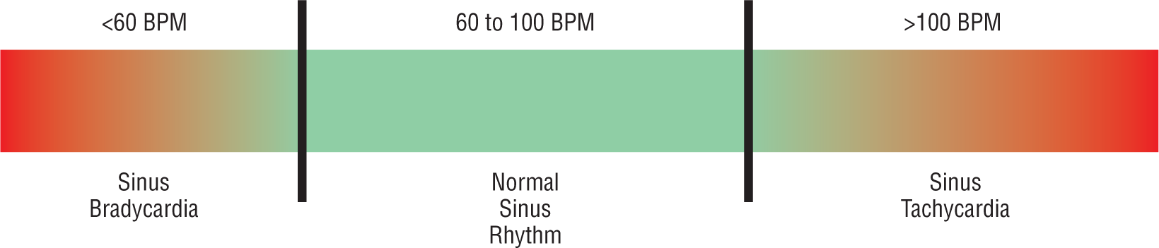
Figure 9-1 The sinus spectrum.
© Jones & Bartlett Learning.
DescriptionSinus bradycardia refers to a rhythm that originates in the sinoatrial (SA) node and has a rate less than 60 beats per minute (BPM). If you look at Figure 9-1, you see that it lies on the left or slower end of the sinus spectrum. Sinus bradycardia has all of the characteristics of normal sinus rhythm (NSR) except that the QRS complexes do not have to be less than 0.12 seconds. The P waves and PR intervals should all be the same morphology and duration.
Figure 9-1 The sinus spectrum.
© Jones & Bartlett Learning.
DescriptionSo, where is the difference between NSR and sinus bradycardia? The answer lies in the phase 4 interval of the cellular action potential (see Figure 9-2 and Chapter 2, Electrophysiology, for a review of the concepts). In sinus bradycardia, the SA node fires slower than in NSR. This means that the automaticity of the cells is prolonged temporally and you end up with a slower and longer phase 4. Electrocardiographically, phase 4 is represented by the TP segment (Figure 9-3). The slower the automaticity, the longer the TP segment, and vice versa.

Figure 9-2 In sinus bradycardia, phase 4 is longer.
© Jones & Bartlett Learning.

Figure 9-3 On the ECG, phase 4 is represented by the TP segment.
© Jones & Bartlett Learning.
Sinus bradycardia is easily identifiable because it has discrete complexes that are usually identical to the NSR complexes but separated by much longer TP segments (Figure 9-3). Once again, notice that the P waves, PR intervals, and QRS complexes are all identical. The QT interval and the duration of the T wave, on the other hand, may be a bit more prolonged in bradycardic rhythms due to their longer repolarization phases.