In Figure 14-1, making the diagnosis of an ectopic P wave was simplified by the presence of an inverted P wave in lead II. Many times, as we saw in Chapter 13, Premature Atrial Contraction, the morphologic difference between an ectopic P and the sinus P is minimal. Take, for example, Figure 14-2. If you were handed this strip, wouldn’t you call it normal sinus rhythm? It meets all of the criteria for normal sinus rhythm, doesn’t it? Clearly making the definitive diagnosis would be quite a diagnostic challenge.
Figure 14-1 This rhythm strip shows the typical changes found in an ectopic atrial rhythm: An ectopic P-wave morphology, rate less than or equal to 100 BPM, normal or slightly prolonged PR interval, and normal QRS morphology.
From Arrhythmia Recognition: The Art of Interpretation, courtesy of Tomas B. Garcia, MD.
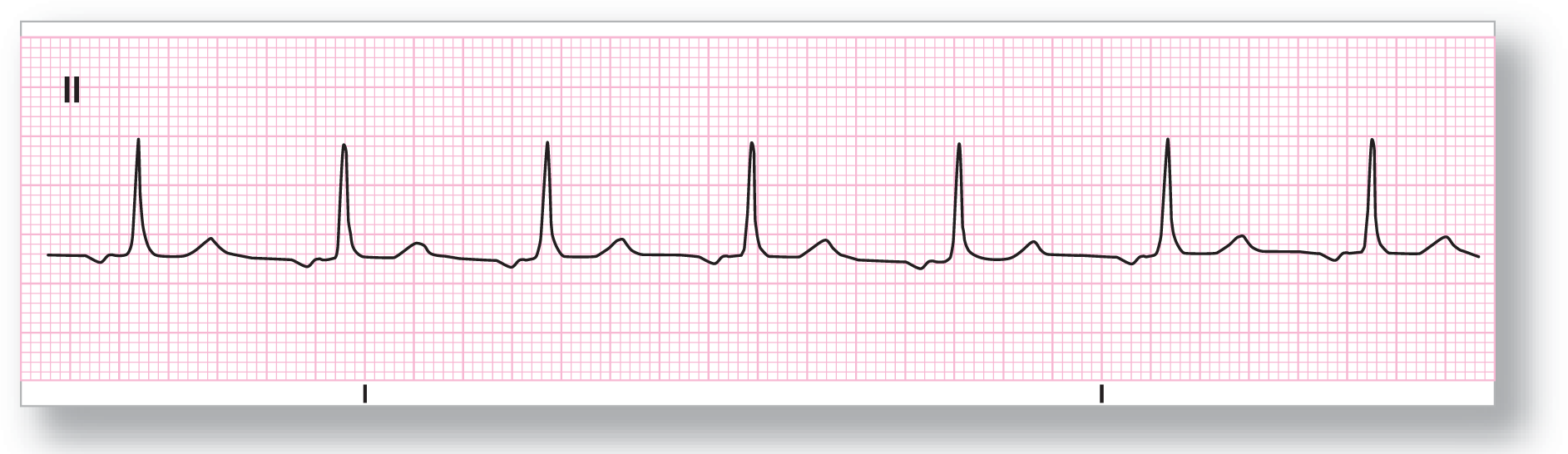
Figure 14-2 This example of an ectopic atrial rhythm would be impossible to correctly diagnose if it were not for the previous strip shown in Figure 14-1. Note the difference in P-wave morphologies and PR intervals.
From Arrhythmia Recognition: The Art of Interpretation, courtesy of Tomas B. Garcia, MD.
In many cases, it is truly impossible to make the diagnosis of ectopic atrial rhythm without some additional clinical or electrocardiographic information. Sometimes, it just takes some plain good old-fashioned luck. However, there are some clues to help you along the way. The rest of this chapter is devoted to those clues and how to recognize them.