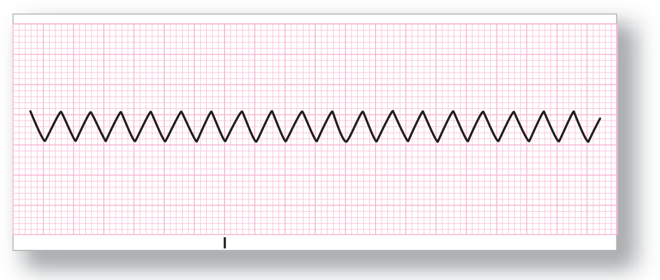
Figure 19-7 Atrial rate at 300 BPM. Note that the peaks fall exactly 0.20 seconds (one big block) apart.
From Arrhythmia Recognition: The Art of Interpretation, courtesy of Tomas B. Garcia, MD.
Now that we have reviewed the concept of AV conduction, let’s take a look at how conduction affects rates. Atrial rates in atrial flutter can vary between 200 and 400 BPM. The most common rate is 300 BPM. Notice that this means that the peaks (either positive or negative) will be exactly 0.20 seconds (one big block) apart on an ECG strip (Figure 19-7). Clinically, that means that if you ever have a strip with a small peak that repeats exactly every 0.20 seconds or one big block, you need to think of the possibility of atrial flutter.

Figure 19-7 Atrial rate at 300 BPM. Note that the peaks fall exactly 0.20 seconds (one big block) apart.
From Arrhythmia Recognition: The Art of Interpretation, courtesy of Tomas B. Garcia, MD.
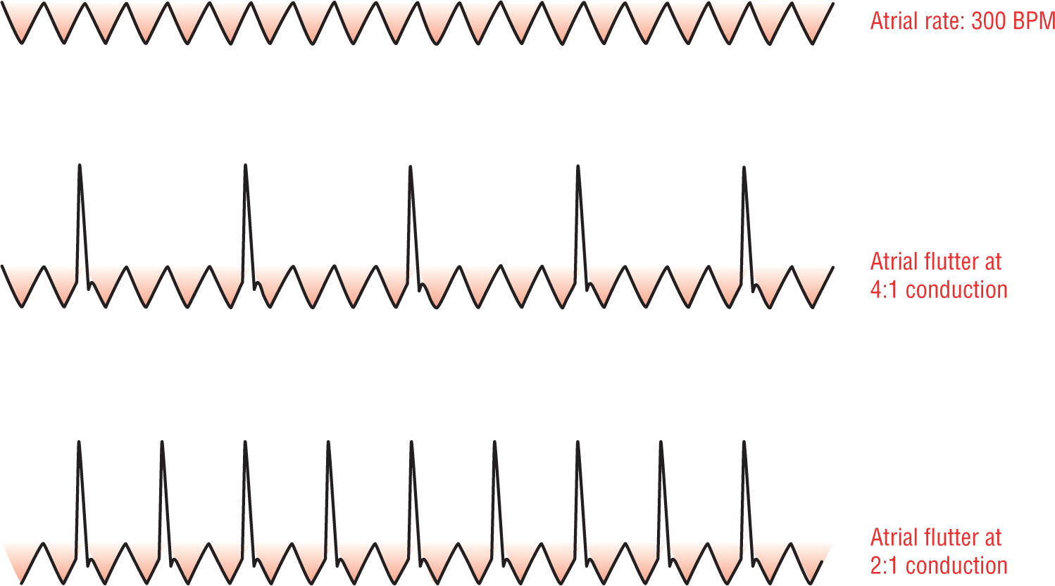
So far we have purposely stayed away from adding in the QRS complexes in order to explain the basic mechanisms involved in the atrial component of atrial flutter. Now, let’s start adding in the ventricular response to the tachycardia (Figure 19-8). The atrial rates (F waves) are typically found around 250 to 350 BPM (and can extend to 200 to 400 BPM in many cases), and the ventricular response ranges from 75 to 175 BPM (most commonly between 140 and 160 BPM). Now, let’s take a huge clinical step: Since the most common atrial rate is 300 BPM and the most common conduction ratio is 2:1, the most common ventricular rate is 150 BPM. Whenever you see a ventricular rate at exactly 150 BPM (or close to it), think of atrial flutter! This is a very important clinical pearl that you need to remember the rest of your clinical life. You will see this come up again and again when you are evaluating rhythm strips and ECGs.

Figure 19-8 Ventricular response in atrial flutter.
© Jones & Bartlett Learning.
DescriptionAdditional Information
Atrial Flutter with Variable Block
The atrial rhythm in atrial flutter is always regular. But, because most AV conduction in atrial flutter occurs at either odd or even ratios (with even ratios being the most common), the ventricular response is usually either regular or regularly irregular.
Regular ventricular responses occur when the ratios remain stable throughout the strip. For example, suppose you had an atrial rate at 300 BPM with 2:1 conduction. The ventricular response would occur regularly at a rate of 150 BPM. Likewise, an atrial rate at 350 BPM with a 3:1 rate would lead to a regular ventricular response.
Regularly irregular ventricular response occurs when the conduction ratios vary throughout the strip (Figure 19-9). An example of this would be an atrial flutter with an atrial rate of 300 BPM with a 3:1 and 4:1 intermittent conduction. When the conduction ratio varies, the regularity varies. But, note that the ratio of conduction is always some multiple of the underlying atrial rate. In other words, it will always be some exact multiple of the distance between the F waves.

Figure 19-9 Regularly irregular atrial flutter.
© Jones & Bartlett Learning.
DescriptionIn addition to the odd and even ratios of AV conduction previously discussed, AV conduction can occur haphazardly throughout the strip, giving rise to an irregularly irregular atrial flutter (Figure 19-10). This type of conduction is usually due to some intrinsic AV nodal disease that does not allow the AV node to function normally. This is an unusual presentation for atrial flutter, but one that you should be aware of because it can easily lead you astray of the correct diagnosis. As we mentioned before, almost all irregularly irregular rhythms are either atrial fibrillation, wandering atrial pacemaker, or multifocal atrial tachycardia. Atrial flutter with variable block is one of those uncommon exceptions to the rule. Note that the term we used was variable block instead of variable conduction, implying that there is some pathologic process at work here.

Figure 19-10 Irregularly irregular atrial flutter.
© Jones & Bartlett Learning.