Chapter 34. Investigating Platyhelminthes, Nematoda, and Annelida
Equipment and Materials
You’ll need the following items to complete this lab session. (The standard kit for this book, available from www.thehomescientist.com, includes the items listed in the first group.)
Materials from Kit
Magnifier
Materials You Provide
Microscope
Slides, prepared, Platyhelminthes
Slides, prepared, Nematoda
Slides, prepared, Annelida
Background
The word worm was applied by early taxonomists to any invertebrate (spineless) animal that was not an arthropod (insect, spider, crustacean, etc.). In common usage, worm is sometimes used to refer to any legless creature with a tubular body, including caterpillars, grubs, maggots, centipedes and millipedes, legless amphibians, and even snakes, none of which are true worms in the original sense of that word. Although taxonomists no longer consider worms a valid grouping, not least because the various species formerly classified as worms do not make up a monophyletic group, the word is still used by biologists as a term of convenience. Worms range in size from microscopic to many meters in length, and can be found in diverse terrestrial and aquatic environments. Some species are free-living predators, while others are parasites that occupy the bodies of various animal hosts.
Like other invertebrates, worms are classified according to the number of structural cell layers they contain and the type of body cavity, if any. The Porifera and Cnidaria we examined in the preceding lab session contain only two structural cell layers, the outer ectoderm and the inner endoderm. As more complex animals, the worms we’ll examine in this lab session contain an additional structural cell layer called the mesoderm, which is located between the ectoderm and endoderm.
In worms, a fluid-filled body cavity may or may not be present in the mesoderm. Worms without a body cavity are called acoelomates. Those with a body cavity that is partially lined with mesoderm are called pseudocoelomates. Those with a body cavity that is fully lined with mesoderm are called coelomates.
In this lab session, we’ll examine some characteristics of representatives of three major worm phyla: Platyhelminthes, or flatworms; Nematoda, or round worms; and Annelida, or segmented worms. These three phyla provide examples of acoelomates, pseudocoelomates, and coelomates, respectively.
Procedure XI-2-1: Observing Platyhelminthes (Flatworms)
As you perform this procedure, record your observations in your lab notebook. Sketch or shoot images of any significant features you observe.
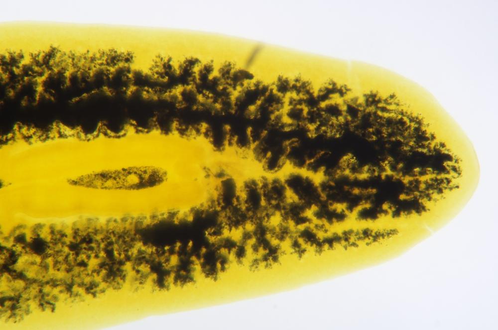
Examine a whole-mount prepared slide of a representative Planarian, such as Dugesia sp., shown in Figure 34-1, with the magnifier and at low magnification with your microscope. Identify any external features visible, such as the pharynx, ocelli (eye spots), auricles (small flaps near the anterior end, used for detecting water currents), and so on.
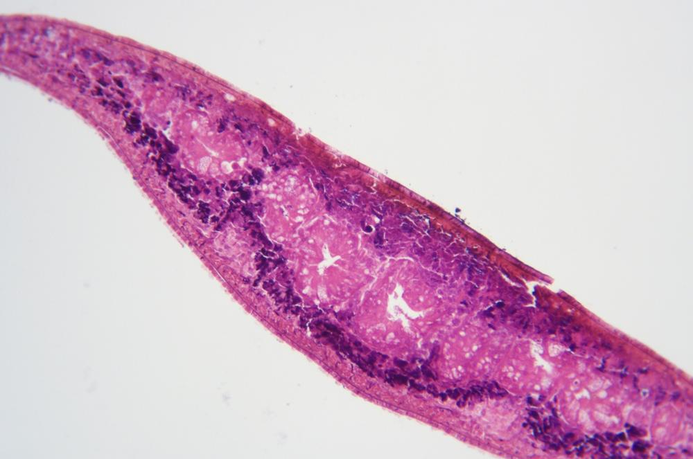
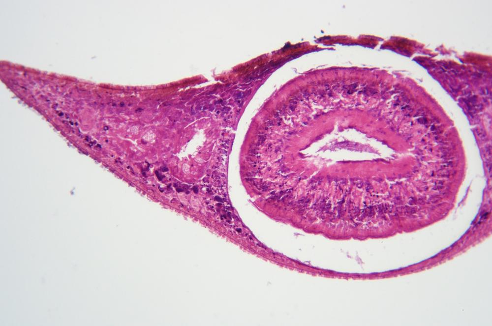
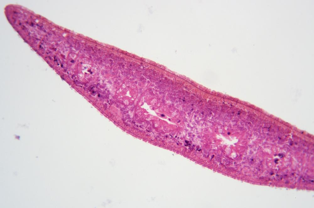
Examine a cross-section of the Planarian at low magnification. Locate and identify the exterior cell layer, the ectoderm, which surrounds and contains the middle cell layer, the mesoderm. The mesoderm is the cellular connective tissue that makes up the bulk of the body, and contains all of the organs. Examine the structures visible within the mesoderm, including the collagen fibers that provide structural support for the body and the muscles that attach to those fibers. Identify the interior cell layer, called the endoderm, a single layer of cells that surrounds the gastrovascular cavity. Figure 34-2 through Figure 34-4 show Planarian cross-sections of the anterior, pharynx, and posterior regions, respectively.
Although Platyhelminthes in the paraphyletic sub-phylum Turbellaria (like the Planaria) are generally free-living and predatory, many flatworm species are parasites that colonize animal hosts and cause serious diseases in humans and animals. These parasites are members of the monophyletic Neodermata sub-phylum, which contains three classes: Cestoda (tapeworms, such as Taenia sp.), Trematoda (flukes, such as Schistosoma sp.), and Monogenea (fish parasites).
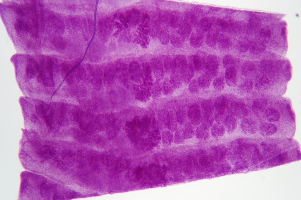
Cestoda are fascinating structurally. Their bodies are made up of a series of segmented plate-like structures called proglottids, which may overlap each other or simply be butted edge-to-edge. The Cestode continues to produce proglottids throughout its life cycle. As proglottids mature, they are expelled from the posterior of the Cestode, mixed with feces. Interestingly, because proglottids contain both male and female reproductive structures, an individual proglottid can reproduce independently. That leads some biologists to consider individual Cestodes to be proglottid colonies rather than single organisms.
If you have prepared slides available, examine whole-mounts and cross-sections of a representative Cestode, such as Taenia sp. or another tapeworm. Compare and contrast the structures with those of the Turbellaria. Identify the proglottids and examine proglottids in all three states: immature, mature, and gravid (filled with fertile eggs). Figure 34-5 is a 40X wm of gravid proglottids from Taenia pisiformis, a tapeworm commonly found in dogs, cats, and other carnivores.
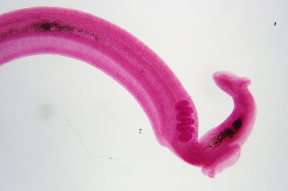
If you have prepared slides available, examine whole-mounts and cross-sections of a representative Trematoda, such as Schistosoma sp., shown in Figure 34-6, or another fluke. Compare and contrast the structures with those of Turbellaria and Cestoda.
Procedure XI-2-2: Observing Nematoda (Roundworms)
As you perform this procedure, record your observations in your lab notebook. Sketch or shoot images of any significant features you observe.
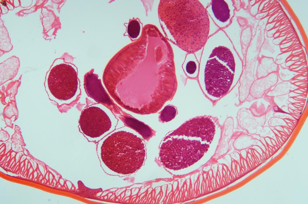
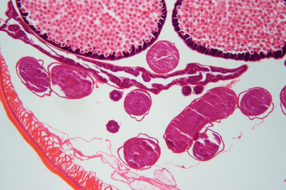
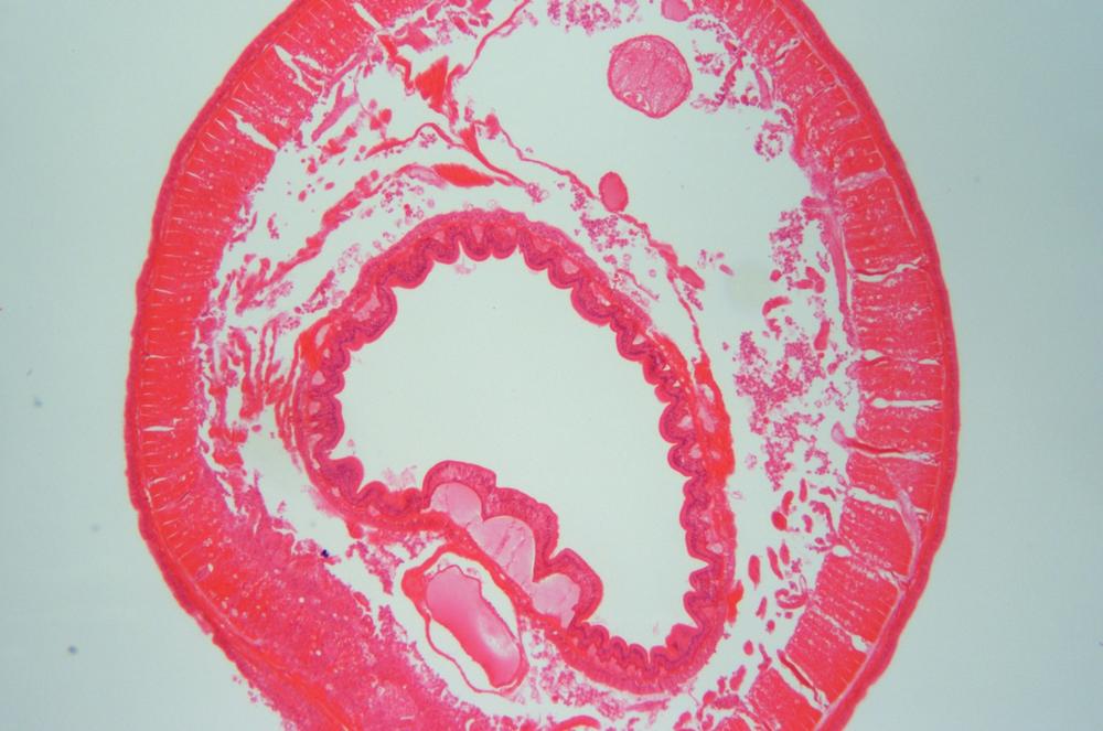
Using low magnification, examine a prepared slide of a cross-section of a representative Nematode, such as Ascaris lumbricoides, shown in Figure 34-7 and Figure 34-8. Working from the outer layers toward the inner layers, identify the following structural features:
- Cuticle
The cuticle is the outermost layer. Depending on the species, the cuticle may be quite thick and have a complex structure, or it may be relatively thinner and simpler. In parasitic species, the impervious cuticle protects the worm against attack by the digestive system or other active defenses of the host.
- Ectoderm
The ectoderm (or epidermis) lies just inside the cuticle, and is a thin single layer of cells.
- Mesoderm
The mesoderm layer lies just inside of the ectoderm, and has a filamentary or fiber-like appearance.
- Pseudocoelom
The pseudocoelom occupies most of the body, from the inner edge of the mesoderm to the outer edge of the endoderm, and is cluttered with other structures scattered throughout it like raisins in a pudding.
- Endoderm
The endoderm is a single layer of cells, bounded on the outside by the pseudocoelom, and surrounding the gastric cavity, which is the digestive tract of the worm and runs its length from the mouth to the anus.
Procedure XI-2-3: Observing Annelida (Segmented Worms)
As you perform this procedure, record your observations in your lab notebook. Sketch or shoot images of any significant features you observe.
Using low magnification, examine a prepared slide of a cross-section of a representative Annelida, such as Lumbricus terrestris, (the common earthworm) shown in Figure 34-9. Working from the outer layers toward the inner layers, identify the following structural features:
- Chaetae
Chaetae (singular chaeta), also called chetae or setae, are thin, stiff bristle-like structures on the exterior surface of Annelids, which provide a grip that allows the worm to move across surfaces, through soil, and so on. The chaetae in Figure 34-9 surround the worm but are barely visible as tiny bumps at 40X. Depending on your specimen, you may have to boost magnification to see any detail in the chaetae.
- Cuticle
The cuticle is the thin, outermost layer, which encloses and protects the inner structures of the worm.
- Ectoderm
The ectoderm lies just inside the cuticle, and is a thin single layer of cells.
- Muscle layers
Lying just inside the ectoderm are two layers of muscles that form the outer boundary of the mesoderm. The thinner outer layer is the lateral muscles, which run around the diameter of the worm; the thicker inner layer is the longitudinal muscles, which run the length of the worm.
- Mesoderm, coelum, nephridia, endoderm, and digestive cavity
The mesoderm layer is bounded on the outer side by the muscle layers, and on the inner side by the muscles that surround the central circular, oval, or horseshoe-shaped digestive cavity. The body cavity between the longitudinal muscles and the digestive cavity is called the coelum. (The true coelum body cavity present in Annelida differs from the pseudocoelum cavity present in Nematoda in that the true coelum is fully lined with mesoderm.) The two filamentary structures, one per side, inside the coelum are called nephridia (singular nephridium).
Review Questions
Q1: If you are given a cross-section slide of an unidentified worm to classify, what single internal feature would allow you to identify unambiguously the worm as Platyhelminthes, Nematoda, or Annelida?
Q2: What characteristic do members of the three phyla we examined in this lab session have in common with each other that they do not share with members of Porifera or Cnidaria?
Q3: Given your knowledge of Annelida structures, propose an explanation for how an earthworm moves forward.