TO understand dinosaurs, we must have some familiarity with their anatomy. In this text, this means skeletal anatomy. A dinosaur skeleton is a complex piece of machinery consisting of more than 300 separate bones, each with its own distinctive features. It is from these bones that paleontologists identify different types of dinosaur and glean much of what we know about these animals. In this appendix, I review the salient features of dinosaur skeletons and the anatomical terms used throughout this book.
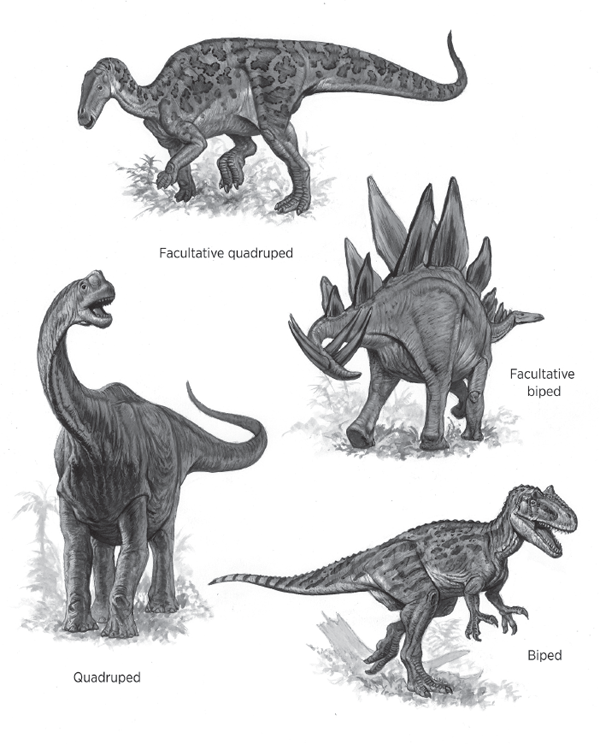
Many dinosaurs were
bipeds, which means they habitually walked on their hind limbs, as do living humans (
figure A.1). Other dinosaurs were
quadrupeds, habitual walkers on all four limbs, such as living horses, cats, and dogs (see
figure A.1). Some dinosaurs walked both ways, sometimes on the hind limbs and other times on all fours. Such dinosaurs were
facultative bipeds (see
figure A.1) or
facultative quadrupeds. Some dinosaurs were facultative bipeds because they walked mostly on all fours but occasionally on their hind limbs, like living bears. Other dinosaurs were facultative quadrupeds because they walked on their hind limbs most of the time and occasionally on all fours, like some living kangaroos.
Whether a dinosaur skeleton belonged to a quadruped or biped or something in between, the terms we use to orient ourselves to it are the same (
figure A.2). The direction toward the head is termed
anterior, and the opposite direction is
posterior. So, paleontologists speak of the forelimbs as anterior to the hind limbs and the tail as posterior to the back. The belly side of a dinosaur is the
ventral side, and the back side is
dorsal. Thus, when we look at a dinosaur skull, the lower jaw is ventral to the eyes, and the nostrils are dorsal to the mouth. Indeed, the top and the bottom sides of a dinosaur skull are referred to as the dorsal and ventral sides, respectively.
FIGURE A.1 Bipeds walk habitually on their hind limbs, and quadrupeds habitually walk on all four limbs. Facultative bipeds and facultative quadrupeds walk part of the time on the hind limbs and part of the time on all fours. (© Mark Hallett. Reproduced with permission of Mark Hallett Paleoart)
FIGURE A.2 Dorsal, ventral, anterior, posterior, lateral, and medial are important terms of orientation in the skeletal anatomy of dinosaurs. (© Scott Hartman)
In any dinosaur, the axial portion of the skeleton consists of the skull, backbone, and tail. In a quadrupedal dinosaur, down and up from the axial skeleton, of course, are ventral and dorsal. But, in bipedal dinosaurs that held their body nearly upright, especially in the front half of the body (such as Tyrannosaurus rex), dorsal points backward and ventral points forward, as in humans. If you are confused when locating dorsal and ventral on a bipedal dinosaur, imagine the animal as a quadruped.
From a horizontal perspective, the direction away from the midline of the body is called lateral. The opposite direction, toward the midline of the body, is medial. Thus, the lungs of a dinosaur are medial to its ribs, but the shoulder blade is lateral to the ribs.
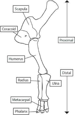
When we speak of dinosaur limbs, however, different terms are used. Those limb segments closer to the body are
proximal to those farther away. And, those limb segments farther from the body are
distal to those closer to the body. So, the knee is proximal with respect to the ankle and, conversely, the ankle is distal with respect to the knee (
figure A.3).
Now we can orient ourselves with respect to any dinosaur skeleton. We can look at the anterior or posterior ends or the ventral, dorsal, or lateral sides. Furthermore, we can locate in a general way any bone with respect to another. A bone is either ventral, dorsal, anterior, posterior, lateral, or medial (or more than one of these) with respect to another bone. And, in the limbs, a segment is either proximal or distal to another segment.
FIGURE A.3 Limb segments farther away from the body are distal to those closer (proximal) to the body. (© Scott Hartman)
Dinosaurs characteristically had an
upright posture (an erect posture). This means the limbs were held directly under the body, as are our hind limbs and the limbs of our pet dogs and cats and most other mammals. This contrasts with the
sprawling posture of most reptiles, in which the limbs are held out to the side so that the proximal bones are horizontal or nearly horizontal to the ground (see
figure 1.2). Some dinosaurs, however, may have held the forelimbs in a semi-sprawling posture intermediate between the upright and sprawling postures.
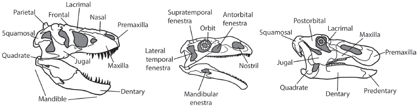
A dinosaur skeleton consists of a
skull, or cranium, lower jaw, or
mandible, and the remaining bones, which are called the
postcrania. The skull of a dinosaur (
figure A.4) is an intricate structure of more than 30 individual bones. These bones are connected to each other along
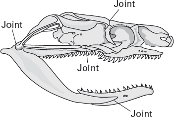
sutures. A sutural connection between two bones is a relatively solid connection and allows little or no movement between the two bones. But, many dinosaurs had joints between various bones in their skulls that allowed these bones to move. The skulls of these dinosaurs are referred to as
kinetic (
figure A.5).
FIGURE A.4 The skull of a dinosaur consists of more than 30 bones, most or all of which are tightly sutured to each other. (© Scott Hartman)
FIGURE A.5 Kinetic skulls, like those of some snakes, have joints that allow skull bones to move past each other. (Drawing by Network Graphics)
Learning the names and locations of all the bones in a dinosaur skull and mandible is not necessary in order to read this book. But, a few key bones should be learned. These bones include the following:
• Premaxillary: the bone at the front of the upper jaw, which in some dinosaurs, bears teeth
• Maxillary: the upper jaw bone behind the premaxillary, which bears the upper cheek teeth
•
Predentary: the bone in the front of the lower jaw, present in some dinosaurs
• Dentary: the principal bone of the lower jaw, it bears the lower cheek teeth
• Jugal: the “cheekbone”
• Parietals and squamosals: two paired bones near the back of the skull
• Frontals and nasals: two paired bones near the front of the skull
• Lachrymals: small bones in front of the eye sockets
• Quadrates: the principal bones of the upper jaw joints
• Occipital condyle: the bony knob at which the skull connects to the backbone
In addition to the names of the individual bones, paleontologists use specific terms to refer to different regions of the dinosaur skull and mandible. The portion of the skull in front of the eyes is termed the rostrum (or face); the eye socket is the orbit; and the region behind the orbit along the side of the skull is the temporal region of the skull. The portion of the skull that encloses the brain is the braincase. The bones outside of the braincase are dermal bones. Openings in these dermal bones in a dinosaur skull are the temporal fenestrae (from the Latin for “window”), consisting of a lower one, termed the lateral temporal fenestra, and an upper one, the supratemporal fenestra. Note that an opening in the mandible is a mandibular fenestra, and an opening in the rostrum in front of the orbit is an antorbital (literally “in front of the orbit”) fenestra. The top of the skull is termed the skull roof, and the area around the nostrils (nasal cavity) is the nasal region. The top of the mouth is the palate, and the lower jaw is often called the mandible.
All the teeth in a dinosaur’s mouth are referred to collectively as its
dentition. Dinosaur teeth come in a variety of shapes and sizes (
figure A.6), but, for all of them, the portion of the tooth above the gumline is called the
crown. When the teeth mesh together, we speak of
occlusion, and the surfaces along which the teeth meet each other are thus the
occlusal surfaces. The crown of a dinosaur tooth may be covered with lophs (ridges) or
denticles (small cusps). When many teeth are present and are attached to (or overlap) each other, they form a
dental battery.
FIGURE A.6 Dinosaur teeth come in many shapes and sizes. (Drawing by Network Graphics)
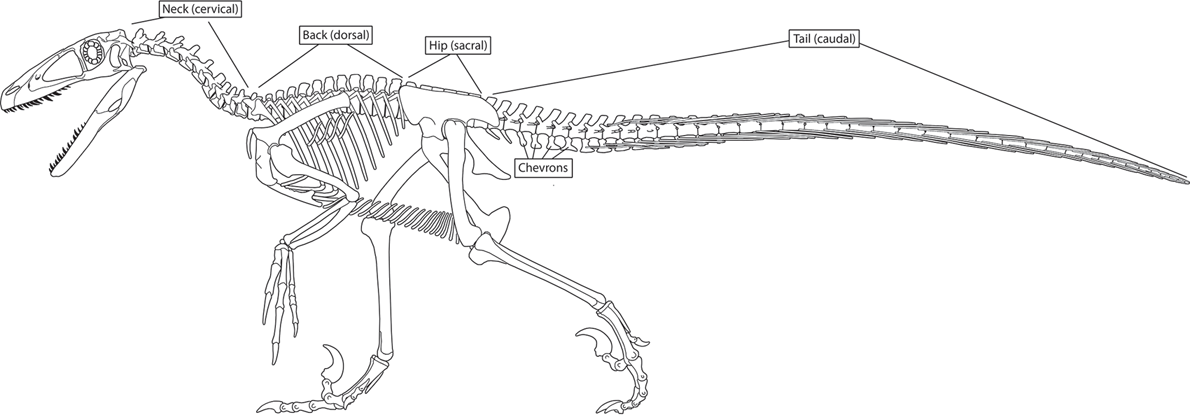
The backbone of a dinosaur is a
vertebral column composed of numerous separate bones called
vertebrae (
figure A.7). Each dinosaur’s vertebral column can be divided into four regions:
cervical (neck),
dorsal (back),
sacral (hips), and
caudal (tail). The number of cervical vertebrae for most dinosaurs is 9 or 10, but hadrosaurs have as many as 15, and some sauropods have as many as 19 cervical vertebrae. Sauropods have an increased number of cervical vertebrae, in part at the expense of their dorsal vertebrae, where they have as few as 9. Most dinosaurs have 15 to 17 dorsal vertebrae and 3 to 5 sacral vertebrae making up the
sacrum, but ceratopsians have as many as 10 sacrals. The number of caudal vertebrae in dinosaurs ranges from as few as 35 to as many as 82 (in the sauropod
Apatosaurus).
Each vertebra has a spool-shaped body along its ventral side, the centrum (plural: centra). Dorsal to the centrum is the neural arch, which covered the spinal cord (it ran through a canal between the centrum and the neural arch). The thin rod or blade of bone that projects dorsally from the neural arch is the neural spine. Ventral to the centrum of the caudal vertebrae of some dinosaurs is another arch-like structure, the chevron. The centra meet each other front to back, and bony ridges link the neural arches with each other, thereby producing an articulated (connected) vertebral column.
Separate ribs attach to the cervical and dorsal vertebrae of dinosaurs. Some dinosaurs also had rib-like bones, the gastralia, covering their bellies.
FIGURE A.7 The vertebral column, composed of many individual vertebrae, can be divided into four regions. (© Scott Hartman)
The forelimb skeleton of a dinosaur attaches to the body at the
shoulder girdle (
figure A.8). The largest and most prominent bone of the shoulder girdle is the
scapula. A smaller bone is the
coracoid. The shoulder girdle in dinosaurs, like ourselves, is held near the front end of the rib cage by muscles and other tissues. Many dinosaurs also had a
clavicle (collarbone) connecting the scapula to the
sternum, a row of bones on the ventral midline of the dinosaur.
The shoulder joint of a dinosaur is the point of articulation between the scapula and the single bone of the upper arm, the humerus. At the elbow joint, the humerus articulates with the two bones of the lower arm, the radius and the ulna.
At the wrist joint, the radius and ulna meet the carpals, the separate bones of the wrist. Distally, the carpals meet the metacarpals, which, in turn, meet the bones of the fingers, the phalanges (singular: phalanx). Pointed terminal (distalmost) phalanges usually bore claws, whereas flattened terminal phalanges bore hooves or hoof-like coverings.
FIGURE A.8 The forelimb skeleton of a dinosaur extends from the shoulder girdle to the phalanges. (© Scott Hartman)
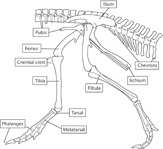
The hind limb of a dinosaur attaches to the body at the
pelvis (
figure A.9). The dinosaur pelvis consists of three bones on each side of the body: the
ilium,
ischium, and
pubis (plural: ilia, ischia, and pubes). Differences in the shapes of these bones, especially in the shape of the pubis, are extremely important in the classification of dinosaurs.
The dinosaur pelvis is securely sutured along the medial surface of the ilia to the sacral vertebrae. The single bone of the upper leg, the femur, fits into a socket, called the acetabulum, at the junction of the three pelvic bones. Distally, the femur articulates with the tibia. The lower leg of a dinosaur also has another, smaller bone, the fibula, which attaches to the tibia.
The tibia and fibula articulate distally with the bones of the ankle, the
tarsals. Two tarsals are important to understanding the origin of dinosaurs. They are the most proximal tarsals, the
astragalus and
calcaneum (see
figure A.9).
Distally, the tarsals articulate with the metatarsals, which, in turn, articulate with the toe bones, the phalanges. As in the forelimb, the phalanges of the hind limb may have borne claws, hooves, or hoof-like coverings, depending on their shape. Fingers and toes of dinosaurs are usually referred to by a single term, digits.
FIGURE A.9 The hind-limb skeleton of a dinosaur extends from the pelvis to the phalanges. (© Scott Hartman)
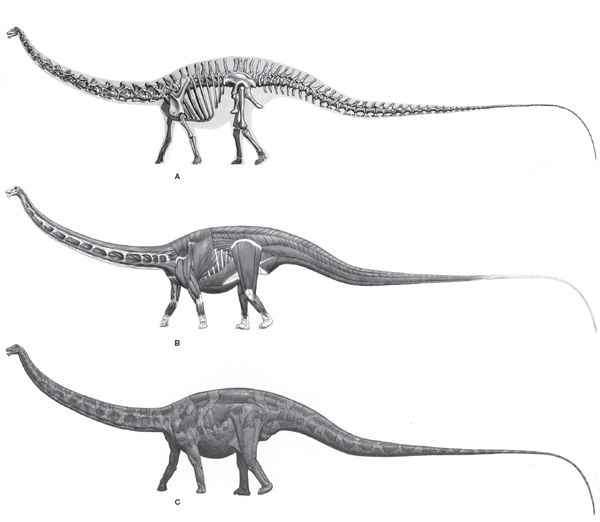
FIGURE A.10 The skeleton of a dinosaur provides the primary basis for interpretations of soft-tissue anatomy and behavior: (A) the skeleton; (B) the same skeleton with muscles restored; (C) the dinosaur as it may have looked in life. (© Mark Hallett. Reproduced with permission of Mark Hallett Paleoart)
This book makes many statements about the behavior of dinosaurs. Some dinosaurs are identified as quadrupeds, others as bipeds. We distinguish plant-eating and meat-eating dinosaurs. Differing ideas about the forelimb postures of some dinosaurs are discussed.
These types of statements reflect the relationship between a particular structure and its
function. This relationship is one of the foundations of paleontology. The size and shape of a single bone, of several bones, or of an entire skeleton depend on, as well as determine, the function of that structure. This is because the skeleton of a dinosaur is the framework to which the muscles were attached and upon which the other soft tissues were hung. How the muscles moved largely depended on the skeleton to which they were attached. And, the shape, size, and arrangement of the other soft tissues—internal organs, blood vessels, and so on—were very much influenced by the skeleton around them. So, it is possible to examine a skeleton and make some inferences about the dinosaur’s muscles and other soft tissues (
figure A.10). This in turn permits an understanding of the appearance and behavior of the dinosaur.
Identification of dinosaurs as bipeds, quadrupeds, or something in between earlier in this appendix and throughout this book provides a good example of the inference of function from structure. Among living animals, bipeds share much larger and more massive (hence stronger) hind-limb skeletons. This makes sense mechanically because the hind limbs of a biped must propel and support its entire weight. In contrast, living quadrupeds have more nearly equally sized forelimbs and hind limbs. Furthermore, the structure of the hips and vertebral columns of living bipeds and quadrupeds differ because of their very different postures while locomoting.
We can look for such structures in dinosaur skeletons as a key to their postures, or more easily just look at relative limb sizes. A clear and easy inference of function from structure can be made by looking at the skeleton of
Tyrannosaurus (see
figure 5.10). Its tiny forelimbs and massive hind limbs indicate bipedality. The more nearly equally sized forelimbs and hind limbs of
Diplodocus (see
figure 6.8), however, indicate quadrupedality.
acetabulum
anterior
antorbital fenestra
articulated
astragalus
axial
biped
braincase
calcaneum
carpal
caudal
centrum (centra)
cervical
chevron
clavicle
claw
coracoid
crown
dental battery
dentary
denticle
dentition
dermal bone
digit
distal
dorsal
facultative biped
facultative quadruped
femur
fibula
function
gastralia
hoof
humerus
ilium
ischium
jugal
kinetic
lachrymal
lateral
lateral temporal fenestra
loph
mandible
mandibular fenestra
maxillary
medial
metacarpal
metatarsal
nasal
nasal cavity
nasal region
neural arch
neural spine
occipital condyle
occlusal surface
occlusion
orbit
palate
parietal
pelvis
phalanx (phalanges)
postcrania
posterior
predentary
premaxillary
proximal
pubis
quadrate
quadruped
radius
rostrum
sacral
sacrum
scapula
shoulder girdle
skull
skull roof
sprawling posture
squamosal
sternum
supratemporal fenestra
suture
tarsal
temporal
temporal fenestra
tibia
ulna
upright posture
ventral
vertebra
vertebral column
1. Orient yourself with respect to a dinosaur skeleton. Relative to the femur, where are the humerus, the maxillary, the tibia, and a caudal vertebra?
2. Compare the segments of the forelimb and hind-limb skeletons, and note any similarities and differences.
3. What is the general relationship between skeletal structure and function?
Dilkes, D. W., J. R. Hutchinson, C. M. Holliday, and L. M. Witmer. 2012. Reconstructing the musculature of dinosaurs, pp. 151–190, in M. K. Brett-Surman, T. R. Holtz, Jr., and J. O. Farlow, eds., The Complete Dinosaur. 2nd ed. Bloomington: Indiana University Press. (A detailed review of how modern muscle anatomy is used to reconstruct dinosaur musculature)
Hildebrand, M., D. M. Bramble, K. F. Liem, and D. B. Wake. 1985. Functional Vertebrate Morphology. Cambridge, Mass.: Belknap Press of Harvard University Press. (An intermediate-level college textbook that examines all aspects of the relationship between vertebrate structure and function)
Hildebrand, M., and G. E. Goslow, Jr. 2001. Analysis of Vertebrate Structure. 5th ed. New York: Wiley. (A college textbook on the inference of structure from function)
Holtz, T. R., Jr., and M. K. Brett-Surman. 2012. The osteology of the dinosaurs, pp. 135–150, in M. K. Brett-Surman, T. R. Holtz, Jr., and J. O. Farlow, eds.,
The Complete Dinosaur. 2nd ed. Bloomington: Indiana University Press. (A review of dinosaur skeletal anatomy)
Romer, A. S. 1997. Osteology of the Reptiles. Chicago: University of Chicago Press. (A detailed technical review of all aspects of the skeletons of reptiles, including those of the dinosaurs; reprint of the 1956 classic)
Romer, A. S., and T. A. Parsons. 1977. The Vertebrate Body. 5th ed. Philadelphia: Saunders. (A thorough review, in introductory textbook form, of all aspects of vertebrate anatomy, including two chapters on the skull and the skeleton)