Whenever a health disorder occurs, it seldom affects only one part of the body. Pain felt in a leg, for example, may originate from pressure on a spinal nerve by a narrowed spinal canal. This, in turn, could be the result of disc herniation. For convenient reading and reference, however, this chapter has been organised into component categories and is based on the format used in the chapter on what can go wrong.
The spinal cord, which is an extension of the brain, occupies the spinal canal within the vertebral column. Anything that places pressure on the tissues or causes injury can produce bothersome symptoms.
Sciatica is an inflammation of the sciatic nerve along its course down the back of the thigh and lower leg.
Symptoms of sciatica may worsen at night or with impending stormy weather, and may last from a few days to several months. They include:
• Mild to severe back pain radiating to the buttock and thigh, worsened by coughing, sneezing or bending
• Numbness or tingling in the lower leg
• Muscle weakness in the buttock, thigh, lower leg or foot
Among the causes contributing to sciatica are:
• Trauma to or compression of the sciatic nerve or its roots, such as that produced by a herniated intervertebral disc
• Inflammation of the sciatic nerve, caused by an infection for example
• Pain referred to the nerve from another part of the body
Increasing the risk of developing sciatica are: a back injury, a herniated disc, being overweight, a sedentary lifestyle and incorrect body mechanics.
• Regular exercise, with emphasis on strengthening the muscles of the back, abdomen and legs.
• Good habits of posture and body mechanics
• Avoiding being overweight
The following are some choices of treatment for sciatica, depending on the symptoms:
• Reduced activity, rather than bed rest, to ease pressure on the nerve and facilitate healing
• Application of heat, such as a heating pad or soaking in a hot bath, to ease pain and muscle spasm
• Analgesic (pain-relieving) or anti-inflammatory medication such as NSAIDs
• Surgery, as in some cases of a herniated disc
• Physiotherapy, such as ultrasound, applications of cold, hydrotherapy and electrical nerve stimulation
• Acupuncture
• Chiropractic (except in cases of a herniated disc)
The word stenosis comes from Greek and means narrow. Spinal stenosis is a narrowing of the spinal canal, which reduces the space available to the spinal cord and nerves.
Those most at risk of developing this condition are: the elderly, people with a herniated disc, spinal tumour, infection or vertebral fracture, and those who are overweight or inactive.
Symptoms of spinal stenosis include:
• Pain in the buttocks and low back, which may radiate to one or both legs
• Numbness in the legs, which eases with rest and bending forwards and worsens with activity
• Weakness and impaired balance
• A foot-slapping gait
Among the conditions that can lead to spinal stenosis:
• Degenerative arthritis
• Bone disease
• Genetic factors
Two of the best measures for helping to avert spinal stenosis are:
• Taking steps to improve posture and body mechanics
• Regularly engaging in simple stretching and strengthening exercises
Additional measures include:
• Controlling your body weight with sensible eating
• Keeping active
• Not smoking (smoking accelerates disc degeneration because it limits blood supply, and it also increases the risk of disc herniation)
NSAIDs are generally useful for reducing inflammation and discomfort.
Other therapeutic measures to consider are applications of heat or cold, and low-impact exercises such as swimming (but first check with your doctor).
For those to whom the above treatments bring no relief, or whose functioning becomes increasingly impaired, injection therapy or decompression surgery may be worth considering.
Although it will not correct the underlying cause, acupuncture may be a useful alternative to other treatments for relieving pain.
Other therapies to try include: ultrasound, diathermy, TENS and massage.
The cauda equina is the terminal portion of the spinal cord. Cauda Equina Syndrome consists of a set of symptoms that characterise a particular disorder.
• Inability to pass urine
• Leaking from a full bladder
• Progressive loss of feeling between the legs
• Progressive loss of power and feeling in both legs
Cauda Equina Syndrome is a result of significant pressure on the structures within the spinal canal. This is a red flag condition, which, because of its serious nature, requires immediate medical attention and possible decompression surgery.
Thirty-three bones form the spine, or vertebral column. Any of these can be damaged by trauma or other forces, including infection, degenerative changes or wear and tear over time.
A fracture, in the simplest of terms, is a broken bone.
Among the symptoms of a fractured spinal bone, or vertebra, are:
• Back pain, sometimes radiating to the buttocks
• Rapidly occurring pain
• Pain while resting
Causes of a fractured vertebra include:
• Trauma, as in a car accident, a sports event or a blow to the back
• Disease, as from osteoporosis and metastases (spread) from cancer (for example, in the breast or prostate gland)
• Use of steroids to treat inflammatory disorders such as arthritis, for example.
If the fracture is due to osteoporosis, certain precautionary measures can be taken to delay its onset or avert its occurrence (see opposite). The chances of fracture from a fall can be lessened by regular exercises that improve balance and coordination and by prudent safety measures, which include sensible footwear and non-skid surfaces in bathtubs and on stairs.
Back pain and associated symptoms occurring after a fall or other accident could indicate serious damage and should be treated as an emergency. The following precautions should be taken:
• Discourage the injured person from moving
• Do not move the injured person
• Call your medical emergency service for assistance (a specially trained team will appropriately mobilise the injured individual’s spine and safely transfer him or her to a treatment facility)
The following are possible measures for treating compression fractures, depending on their cause:
• An extension brace, to be worn for about three or four months, with periodic X-rays taken to assess healing or deformity
• Good posture when lying down, avoiding the prone position
• Possibly taking nutritional supplements to promote bone health, such as calcium and magnesium, boron, zinc and vitamin D (check with a qualified nutrition specialist)
• Avoiding alcoholic, caffeinated and carbonated drinks, which can detract from bone health
• Analgesics to relieve pain, and anti-inflammatory drugs to counteract inflammation
• Fairly rapid but graduated mobilisation following impaired functioning, to build strength and confidence (a physiotherapist may be helpful)
• Appropriate treatment if osteoporosis is the cause of fracture (see opposite)
When less invasive treatments fail to bring the desired results, surgery may become necessary. Operations include vertebroplasty and kyphoplasty.
Osteoporosis, which literally means ‘porous bones’, describes a disease process in which bones become brittle and weak and prone to fractures. The most serious loss of bone integrity occurs in the spine and femur (thigh bone). Once weakened, the vertebrae can become easily compressed by the weight of the body itself. When this happens, it can reduce your height by several inches/centimetres.
Difficulty in bending forwards and back pain are two symptoms that suggest osteoporosis. As the disease process advances, height may be noticeably decreased, and sometimes a kyphosis (dowager’s hump) may develop. If the bones of the ribcage are involved, it may affect efficient breathing.
Osteoporosis mostly affects post-menopausal women and, to a lesser extent, sedentary men. It can also occur among young women who drastically limit their food intake to become or stay thin: nutrients crucial to the health and strength of bones, such as calcium, vitamin D, boron and zinc, become inadequate.
Bone integrity can also be compromised by the frequent use of antacids (substances that neutralise acid) containing aluminium, since they accelerate the excretion of calcium. Some diuretics (water pills), which promote calcium loss, and some laxatives, especially mineral oil, if used frequently, can deplete supplies of important mineral and vitamins. In addition, steroids, sometimes administered in the treatment of inflammatory diseases such as arthritis, may inhibit bone formation and calcium absorption.
Preventive measures should begin in early adulthood to build bone mass and strength. This includes a diet rich in bone-building nutrients such as calcium, vitamin D, zinc and boron and regular weight-bearing exercise such as walking, dancing and stair-climbing.
It would be prudent, in addition, to avoid smoking, which is believed to be toxic to bone, and to keep consumption of alcohol, caffeinated beverages and carbonated drinks low, since these are thought to deplete the body of essential nutrients. Avoid regular use of antacids containing aluminium, which tend to promote bone loss.
Learn and habitually practise stress-management techniques to help keep the level of cortisol (stress hormone) low.
Once osteoporosis has occurred, treatments tend to produce less than ideal results. Nevertheless, ensuring the intake of essential nutrients, such as those already mentioned above, is useful in slowing down the progression of bone loss. Best among the food sources of such nutrients are: green leafy vegetables, dairy and soybean products and whole grains. Nutritional supplements may also be helpful, but first check with your doctor or other qualified health professional.
Post-menopausal women should discuss hormone replacement therapy with their doctor, and possibly also other medicines to increase bone mass and decrease bone loss.
Regular exercise is essential not only in helping to prevent osteoporosis but also to slow down its progression. Caution is needed, however, in attempting to do flexion exercises (forward-bending movements) if the vertebrae are involved. Please check with your doctor or physiotherapist. Exercises to improve coordination and balance are important, as they can help to prevent falls and possible resulting fracture.
Stress management strategies should be part of daily life, as mentioned above. Medicines used to treat osteoporosis include:
• Bisphosphonates, which reduce bone resorption (loss of bone substance), and which may also increase the activity of bone-building cells (osteoblasts)
• SERM (Selective Estrogen Receptor Modulations), which reduce bone resorption, but which should be given only post-menopausally
• Calcitonin to reduce bone resorption and possibly increase bone-building cells, and which may also be useful in relieving pain
• Teriparatide (Forteo), an injectable hormone derived from the parathyroid glands (which are involved in calcium metabolism), which helps to increase bone formation
• Hormone replacement therapy may be considered to increase osteoblast activity and reduce bone resorption, but it may not be appropriate for all women. It should be carefully discussed with your doctor, as it has the potential to produce some adverse effects.
• Non-prescription analgesics such as aspirin and paracetamol can provide pain relief.
• Local applications of heat, such as a heating pad
• Nutritional therapy
• T’ai Chi
• Yoga
Joints are connections between bones. Between every two vertebrae are joints formed by smooth cartilage and strengthened by ligaments that run behind and in front of the vertebral bodies throughout the entire length of the spine. In addition, there are facet joints at the back of the vertebrae. Like joints elsewhere in the body, spinal joints are vulnerable to injury by infection or tumour, by degenerative processes, or by trauma.
Known also as non-inflammatory arthritis or degenerative joint disease, osteoarthritis is the most common type of arthritis and tends to occur in older individuals. It affects the smooth cartilage covering the ends of bones where they form joints. It can also involve the underlying bone and surrounding tissues and other spinal components, such as discs and ligaments.
Although inflammation is not typical of OA, changes in the joints may sometimes produce a local inflammatory response.
Stiffness and back pain, which may worsen with extension (bending backwards), could be an indication of spinal osteoarthritis. The pain, however, is rarely disabling.
OA in the low back can lead to a narrowing of the spinal canal (see stenosis) or to slippage of a vertebra (see spondylolisthesis, page 000) and produce other symptoms, including pain that radiates to, and numbness and weakness in, the legs.
OA can develop following trauma or other injury, an inflammatory joint disease, or repetitive strain related to your occupation or sports activities.
To help prevent the occurrence or progression of OA:
• Maintain good general health, with attention to a diet rich in essential minerals and vitamins
• Avoid being overweight
• Exercise daily to maintain spinal strength and flexibility
• Practise good posture and body mechanics to minimise stress on spinal structures
Among the treatment choices for dealing with OA are:
• Heat, through local application such as a heat pack, by immersion in a whirlpool bath, or with a warm shower
• Hands-on treatments such as Shiatsu or other form of massage (inflamed areas should not be massaged, however)
• Non-prescription analgesics such as paracetamol
• Topical application of capsicum cream. (Capsicum is found in chilli, red and cayenne peppers and has anti-inflammatory properties.)
• Stretching and strengthening exercises
• Injection therapy or surgery where applicable
• Acupuncture for pain relief
• Herbal medicine, including sources of essential fatty acids (EFAs, see below) to help reduce inflammation
• Nutritional therapy, which includes supplements of nutrients such as vitamins C, D and E, and also of glucosamine (a cartilage-building material and a component of synovial fluid) and chondroitin (a vital part of the 'glue' that maintains the integrity of joint cartilage)
• Yoga and T’ai Chi
• Relaxation techniques
EFAs are called ‘essential’ because the body must have them to carry out certain vital functions. But since the body cannot make them, they have to be provided by food. Food sources of EFAs include flax seeds, soy beans and walnuts, and oils from these; dark green leaves; and cold-water fish such as salmon, sardines and trout. Supplementary sources of EFAs include oils from blackcurrants, borage and evening primrose.
Rheumatoid arthritis is an inflammatory type of arthritis that affects connective tissue, most notably synovial joints (which contain a lubricating fluid to facilitate movement). Its incidence is two times higher in women than in men, until the age of 65 years, when both are almost equally affected.
For most RA sufferers, the condition first produces fatigue and general malaise. Stiffness is noticeable after inactivity, especially after sleeping at night. With progression of the disease, loss of function increases.
Although RA affects mostly the hands and feet, it also occurs not infrequently in the cervical spine. If neck involvement is severe, spinal cord compression may result and surgery may become necessary.
RA can develop in the low back also, where it can affect facet joints, ligaments and other adjacent structures and produce pain.
RA was once believed to be an autoimmune disease, in which the body literally turns against itself, but this is currently in question.
There appears to be a genetic predisposition to RA, and hormonal fluctuations seem to play a part also. For example, symptoms tend to abate during the last trimester of pregnancy but flare up following childbirth. Infection can also be a contributing cause.
Preventive measures to take against RA include joint protection and regular appropriate exercise to help to maintain function. Joint protection consists essentially of learning and practising good habits of posture and body mechanics and taking breaks from prolonged or repetitive activity to do simple stretching exercises. Regularly engaging in appropriate exercises is one key to maintaining function and preventing disability. A physiotherapist or other qualified professional can help you. The therapist can also be of assistance in the safe use of mobility aids such as walkers.
Treatments for people with RA aim to relieve pain, reduce inflammation, protect joint surfaces and maintain function. In addition to relieving pain and reducing inflammation, anti-inflammatory medications are also used to help prevent the progression of RA from a localised area to other body parts. These drugs include aspirin, NSAIDs such as ibuprofen and naproxen to decrease pain and swelling, short-term use of glucocorticoids such as prednisone and cortisone, and slow-acting anti-rheumatic drugs.
• Local applications of heat
• Massage therapy
• Nutritional supplements including EFAs (essential fatty acids), glucosamine sulphate and chondroitin sulphate (see Osteoarthritis)
• T’ai Chi
• Yoga
• Relaxation techniques
Ankylosing spondylitis is a chronic, progressive inflammation of the spine and sacroiliac joints. It affects mostly males, usually before the age of 40 years. Its origin is unknown, but a strong hereditary tendency is suspected.
The most common symptom of AS is pain in the low back and upper buttocks, which comes on gradually and persists for at least three months. It worsens with rest and eases with graduated activity. The pain can also radiate to the upper back and sometimes to the breastbone. Stiffness, which is more noticeable after a night’s sleep, is another symptom.
AS can also affect other body tissues, including those of the heart, lungs, kidneys and eyes, spinal ligaments and tendons, sacroiliac joints and the joints between the ribs and vertebrae, which can cause breathing difficulties.
Other manifestations of AS include general malaise and weight loss and, as the disorder progresses, a fusing together of the vertebrae, increased arching of the upper back and rigidity of the neck.

The cause of AS is not known with certainty, but heredity and infection are thought to be implicated.
There appear to be no effective measures that can prevent or slow the progress of AS. Goals of treatment are: to relieve pain through the use of NSAIDs and heat, for example; to maintain the best possible posture, if necessary with the help of a back brace or splints, so as to prevent breathing problems, and taking short periods of rest to counteract fatigue and maximise energy reserves.
Appropriate exercise to help maintain strength and mobility is advised.
Surgery is rarely necessary to correct deformity of the lumbar spine.
Facet joints are paired synovial joints at the back of the vertebrae. The capsules of the joints are richly supplied with nerve endings and pain fibres that are sensitive to strain, pressure and stretching.
Like other joints, facet joints are prone to injury and are a not uncommon source of low back pain
Pain both local and referred, muscle spasm and tenderness, and reduced spinal flexibility are the chief symptoms of facet joint injury. Pain is usually worse on extension of the spine and less with flexion.
Any force that stretches the capsule surrounding a facet joint, and also the ligaments, beyond their tolerance can induce an inflammatory response, resulting in joint pain and swelling.
• Regular practise of stretching and strengthening exercises
• Frequent breaks from repetitive work to do tension-relieving exercises
• Posture training, such as that provided by the Alexander Technique
Anti-inflammatory medications such as NSAIDs and aspirin may be taken to relieve pain and inflammation.
In selected cases, injection of a steroid such as cortisone directly into the affected joint may be an option. This treatment may be useful, for example, when the symptoms are produced by an inflammatory arthritis. One or two repeat injections may be necessary. The efficacy of this therapy for facet joint pain is controversial, however.
From the Greek spondylos, vertebra, and olisthanein, to slip, spondylolisthesis refers to a condition in which one vertebra slips over another. This most commonly occurs between the fifth lumbar and the first sacral vertebrae.
Although the spinal instability caused by spondylolisthesis in adults may produce no symptoms, it can sometimes result in back pain. When the condition causes a stenosis, or narrowing in the spinal canal, it can generate symptoms of nerve compression (see spinal stenosis).
Arthritic changes in the lumbar spine, most common in older women, are one cause of spondylolisthesis. This can result in a narrowing of the spinal canal.
This condition is also sometimes seen in adolescents and may cause back pain, tight hamstring muscles and nerve irritation.
Preventive measures include:
• Regular practise of exercises to strengthen the back and prevent tightness of the hamstring muscles
• Careful attention to posture, body mechanics and correct technique in sports
• Avoiding being overweight
Spondylolisthesis due to degenerative changes is treated along lines similar to those for spinal stenosis and for osteoarthritis. Other treatments include:
• Appropriate stretching and strengthening exercises
• Postural education
• Avoiding becoming overweight
• Not smoking
• Nutritional therapies
• Anti-inflammatory medicines, if necessary
• Injection therapy, sometimes
Surgical treatments, when considered necessary, consist of decompression and spinal fusion.
Between the weight-bearing parts of vertebrae, from the second cervical to the sacrum, are intervertebral discs. They consist of an outer fibrous ring (annulus fibrosus, or AF) and a soft inner substance (nucleus pulposus, or NP). For more details. Although the discs can withstand tremendous compression, they do not tolerate rotational stress very well. To compensate, the facet joints help to resist excessive twisting forces.
Disc degeneration is a rapid progression of changes in the discs that result in a loss of their integrity and in their ability to distribute load.
Degenerative changes in the spinal discs should not be equated with ageing. Although all discs age, they do not necessarily degenerate.
Degeneration of intervertebral discs does not always produce symptoms. When it does, it can cause a great deal of pain, which worsens with flexion.
Included in the possible causes of DDD are:
• Excessive force applied to vertebrae through heavy lifting, for example
• Cracks and tears in the AF and leaking from the NP (see above)
• Injury to the discs by excessive pressure, which may be aggravated by rotational forces. The combination can result in an AF tear, which in turn can induce disc herniation
Preventive measures include:
• Using correct techniques when exercising and lifting heavy objects
• Using appropriate protective gear when working with vibrating equipment
• Taking periodic relaxation breaks when driving long distances
• Avoiding being overweight
As for many other disorders of the spine, therapy for DDD should include:
• Stretching and strengthening exercises
• Training in good posture and body mechanics
• Losing weight if necessary
• Not smoking
• Where appropriate, injection therapy and surgery may be considered.
A weakness in the AF of an intervertebral disc may permit a bulging of the NP; a break or tear allows the NP to protrude or push outward from its normal position. These occurrences, which have given rise to the misnomer ‘slipped disc’, refer to a herniated disc.
Disc herniation is common among males between the ages of 30 and 50 years and can appear within 36 hours of the precipitating cause.
Among the symptoms occurring with disc herniation are:
• Pain in the back, worsened by flexion and eased by extension
• Pain, numbness and weakness in the legs
• Muscle spasm
• Symptoms suggestive of Cauda Equina Syndrome
An entrapped cervical disc undergoes a process similar to that of a herniated disc in the lumbar spine. Symptoms may include neck pain and spasm, changes in sensation in the hand, and a weakened hand grip.
Changes occurring with the passage of time in both the AF and NP components of spinal discs can contribute to disc herniation.
A fall or other accident, an injury from twisting or lifting, or even an episode of coughing or sneezing can precipitate disc herniation.
Sensible preventive measures for minimising the risks of a herniated disc include:
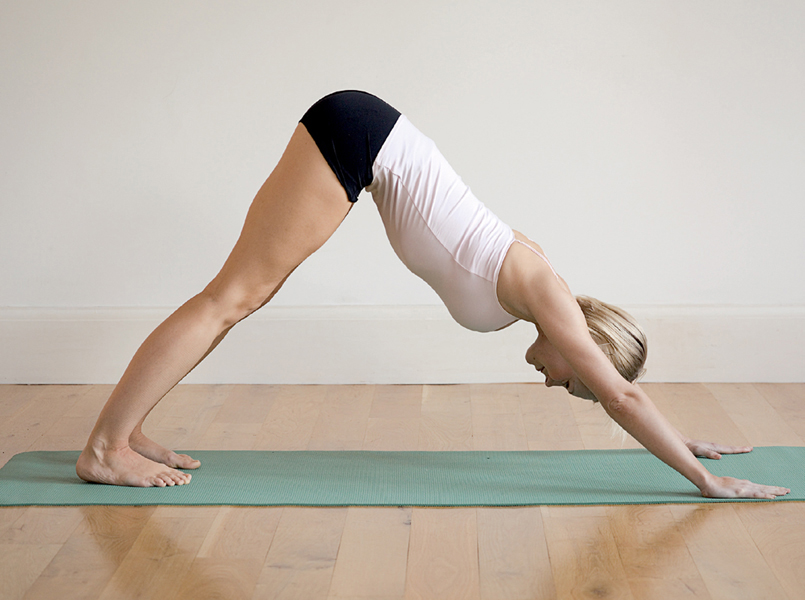
• Regularly engaging in exercises that strengthen the torso, keep the spine flexible and improve balance and coordination
• Taking periodic short breaks from repetitive work to prevent tension build up and unnecessary strain
• Maintaining proper posture at work and elsewhere, to reduce stress on the spine and its components
• Avoiding being overweight
• Wearing proper-fitting low-heeled shoes, to minimise strain on spinal structures
Included in the treatments for acute disc herniation, where no red flag symptoms appear, are the following:
• Restricted activity but no bed rest, except in severe cases, and then not beyond 48 hours
• Applications of heat to the affected area, to ease muscle spasm and pain
• Medications such as analgesics and anti-inflammatory agents, and cautious use of narcotics
• Gentle exercise (excluding flexion exercises) in consultation with a physiotherapist or other qualified health professional, balanced with periods of relaxation
• Good habits of posture and body mechanics
Steroid injection, discectomy and chemonucleolysis are three minimally invasive options to consider in selected cases.
For a mild to moderate cervical disc problem, a soft cervical collar may be prescribed to limit neck mobility.
During the acute phase of disc herniation, chiropractic and massage are not recommended.
• Acupuncture may be considered for pain relief
• Strategies to help stop smoking would be prudent since smoking is believed to be injurious to the health of the discs
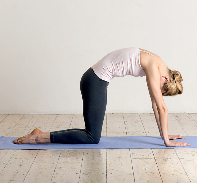
• Yoga and T’ai Chi, which are excellent in helping to attain and maintain flexibility, balance and coordination, are two exercise approaches to consider
• For posture education, the Alexander Technique is superb
When viewed from the side, the normal spinal column shows four curves. These curves contribute to the strength of the spine, absorb shock, help us to maintain balance and give a measure of protection from fracture.
In a normal spine, the lumbar curve bulges forwards. When it is exaggerated, it is known as lordosis.
Any departure from the normal condition of one of the body’s structures could affect adjacent tissues. Lordosis subjects other spinal components to muscular strain and may therefore lead to fatigue and pain. If spinal discs become involved, additional symptoms may appear.
Poor postural habits over time can lead to lordosis. The habitual wearing of high-heeled shoes may also be a contributing factor since it alters the body’s centre of gravity. Lordosis is also often seen in pregnancy, for the same reason.
Obesity or a large abdominal tumour can also produce this abnormal spinal curvature.
Two keys to preventing lordosis are:
• Good habits of posture and body mechanics
• Maintaining the strength of the back and abdomen through regular exercise
• Wearing sensible shoes is an additional prudent preventive measure to take (see also Orthotics).
Prevention is the best treatment for lordosis. In some cases, however, the temporary use of a brace may be required, and in a few selected cases the extreme measure of spinal fusion surgery may be considered.
Kyphosis, also referred to as humpback or dowager’s hump, is the result of excessive curvature of the thoracic spine.
Like other posture defects, kyphosis can cause fatigue and back pain. A severe curvature that affects the functions of the heart, lungs, stomach and intestines can produce symptoms including an irregular heartbeat, difficulty in breathing and problems with digestion and elimination.
Kyphosis is common in metabolic disorders such as osteoporosis and osteomalacia (softening of bones). It is also sometimes associated with muscular dystrophy, a condition in which muscles weaken and waste away.
To help prevent posture defects from developing:
• Exercise regularly to keep limbs and torso strong and flexible
• Posture training and practising good habits of posture and body mechanics are crucial
• Maintaining the weight that is ideal for you is a prudent measure to take
• An exercise regime to strengthen muscles and ligaments
• Braces may be used to help improve the deformity
• Analgesics or anti-inflammatory medication can be used to relieve pain and symptoms of inflammation
• If the kyphosis is caused by osteoporosis, appropriate therapeutic measures should be taken
• If response to these therapies is unsatisfactory, kyphoplasty or other surgery may be required
• Physical therapies including applications of heat and cold, ultrasound, electrical nerve stimulation and massage
• Alexander Technique or other methods of posture training
• Yoga or T’ai Chi for exercises in stretching, strengthening, balance, coordination and relaxation
• Chiropractic for corrective manipulation and strengthening exercise
Scoliosis is lateral (side-to-side) curvature of the spine in any area. The most common form appears in adolescents and preadolescents.
Apart from possible backache and fatigue resulting from the muscular imbalance caused by scoliosis, breathing difficulties and heart irregularities can appear if the ribcage is seriously deformed.
Scoliosis can be present at birth (congenital) or due to neuromuscular (nerve-muscle) conditions or spinal trauma.
Regular monitoring, preferably by an orthopaedic specialist, is useful in detecting any worsening of the condition.
Non-surgical treatments include:
• Exercise, including swimming and horseback riding to strengthen torso muscles and promote good posture.
• The use of braces for several hours a day may be advised, according to individual requirements
• A physiotherapist can help you organise having the heel of one shoe built up to equalise the length of the legs
• Where necessary, appropriate treatment for the condition producing the spinal defect is given
The decision to proceed with surgery is based not only on the ineffectiveness of conservative treatments, but also on whether the patient is willing to undergo a major operation.

Ligaments are bands of fibrous tissue that hold bones together. Tendons are fibrous bands that attach muscles to bones. Both can be injured by overstretching, overuse, trauma and poor posture and body mechanics.
A sprain is a traumatic injury to the ligaments, tendons or muscles around a joint. In the spine it often affects the ligaments that bind together the facet joints. This occurs most often in the lumbar region, but the sacroiliac joints are sometimes involved.
A sprain usually produces pain, swelling and discolouration of the skin over the affected joint. The pain sometimes radiates to the buttocks and legs. The duration and severity of these symptoms vary with the extent of damage to the supporting tissues.
Torn or injured ligaments are usually the result of a fall or an activity that produces a wrenching action. They can also be due to overuse, as sometimes occurs in gymnastics or ballet, and from the long-term impact of poor posture and body mechanics.
During pregnancy, when ligaments become lax, some women may be vulnerable to sacroiliac sprain.
Measures to aid the prevention of sprains include:
• Regular exercise to keep the body strong and flexible
• Frequent breaks from repetitive work to do tension-relieving exercises and so avert cumulative strain
• Good habits of posture and body mechanics
• Warming up adequately before engaging in sports and other vigorous activities
These include:
• A support device such as a brace, if necessary
• Applications of ice or alternating this with heat
• Ultrasound treatment to help speed up recovery
• Short rest periods, but no prolonged bed rest (which tends to weaken muscles, discs and cartilage, worsen existing back problems and accelerate bone loss)
Muscles supporting the spine can be vulnerable to a number of injuries including tears, strain and cumulative stress, and also to the development of hard knots called ‘trigger points’.
Although strain can occur at any level of the spinal column, its most frequent site is the lumbar area. Sometimes the condition is referred to as ‘myofascial strain’ (myo = muscle; fascia = surrounding tissue).
Pain is one of the first symptoms of back strain and can range in intensity from mild to severe. The pain, which may radiate to the buttocks, usually appears within 36 hours of the precipitating cause, often an injury caused by a fall, a twisting action, or by the lifting of a heavy object. It is aggravated by movement and relieved by rest, and can be worse on flexion of the spine. Muscle spasm may also be present, and there may be localised swelling and tenderness.
Back strain is usually the result of a minor injury incurred by a fall, a twisting motion, or by incorrectly lifting an object.
Keys to helping prevent back strain include:
• Regular practise of stretching and strengthening exercises
• Good habits of posture and body mechanics
• Frequent breaks from repetitive work to practise stress-relieving exercises
• Controlling body weight through sensible nutrition and exercise
In the acute stage, back strain in the lumbar area is best treated with:
• Ice and restricted activity (limited bed rest; gentle stretching exercises and walking)
• Attention to good posture
• Analgesics or anti-inflammatory medicines, if appropriate
Not recommended at this stage are:
• Application of heat
• Massage
• Braces and other supports
These include: TENS and acupuncture to help speed up rehabilitation, and chiropractic in the absence of radiating pain (as in sciatica with numbness and weakness).
Provided that red flag symptoms are absent:
• Ultrasound or local applications of heat to reduce spasm, improve function and promote healing
• Ice, following stretching or other suitable exercise
• Prudent use of analgesics or anti-inflammatory medications, with decreasing frequency
• Acupuncture
• Hands-on therapies including Shiatsu or other form of massage
• Yoga or T’ai Chi to promote strength, flexibility, balance and coordination
As mentioned at the start of this chapter, whenever a health disorder occurs, it seldom affects only one part of the body. The neck, or cervical spine, is an example: injury can affect any or all of its bones, joints, nerves, discs, ligaments or muscles. Making the neck even more vulnerable is the fact that it is usually very flexible and capable of a wide range of motion.
Whiplash is a word used to describe certain neck movements under specific conditions. It is a mechanism rather than an injury or illness. It may or may not cause injury.
These vary according to the degree of injury involved. A whiplash movement may produce no symptoms or outward signs of mishap, or it may result in any of the following: neck stiffness and a decrease in the neck's range of motion; tenderness and pain; evidence of neurological injury such as altered sensation and muscle weakness along with pain, or fracture or dislocation of cervical vertebrae, revealed by X-ray. There could also be feelings of dizziness, ringing in the ears and pain in the jaw or arms.
In addition, pain in the cervical spine can spread to the head and produce a headache. This is because the skull is covered by a sheet of muscle that extends from behind it, near the upper neck, to the eyebrows (occipito-frontalis). Tension in the neck can be transmitted to this muscle, which then tightens – much like a drawstring – and can generate a headache.
Whiplash injury is most often caused by a rear-end car crash that causes the neck of the driver or passenger to be jerked too far backwards (hyperextend) and then too far forwards (hyperflex) and sometimes forwards again. If the vehicle crashes into a stationary object or is involved in a head-on collision, the sequence can be reversed.
Although accidents cannot always be prevented, certain measures can be taken to minimise their impact. These include:
• Keeping fit through regular conditioning exercises to keep the body strong, flexible and well toned
• Good habits of posture and body mechanics
• Avoiding being overweight, in order not to stress various body parts unnecessarily
Airbags and properly designed headrests also offer some protection.
Injury resulting from a whiplash movement could cause fracture and/or dislocation of a vertebra with resulting compression of the spinal cord. This is a medical emergency for which professional help should be summoned immediately:
Choices of treatment for whiplash injury depend on the individual case. Generally they include:
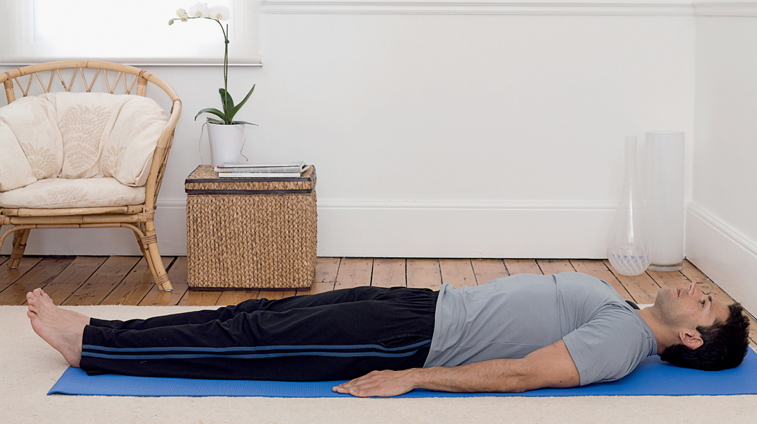
• Short rest periods while lying on a firm supportive surface
• Gentle neck-stretching exercises for the neck and shoulders as tolerated (check with your doctor or physiotherapist)
• Application of ice alternated with heat, in a 20-minutes on and 20-minutes off cycle
• Pain-relieving or anti-inflammatory medicines as required
• Treatments given by a physiotherapist including: whirlpool baths, hot and cold packs, electrical nerve stimulation, diathermy, ultrasound or gentle massage
• Taking short, frequent breaks from prolonged sitting (at a desk or computer, for example) to prevent cumulative tension and fatigue
• Surgery may be necessary in some cases of vertebral fracture or dislocation
• Alexander Technique or other posture training method
• Chiropractic or osteopathy in selected cases only (not where osteoporosis is present)
• Relaxation techniques (inherent in methods such as Autogenic Training, biofeedback and yoga)
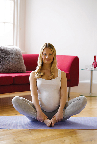
Backache in pregnancy is a common occurrence and is largely due to a change in the woman’s centre of gravity and to the effects of a hormone called relaxin. Relaxin literally relaxes ligaments that support joints, particularly where the spine meets the sacral bones (sacroiliac joints). This is one of nature’s ways of facilitating an increase in the size of the passageway to accommodate the child’s birth.
As pregnancy progresses and the weight that the woman carries in front increases, her centre of gravity is altered. To compensate, the back muscles have to work harder to help her maintain balance. The pelvis tilts farther forwards and this increases the curve of the lumbar spine (lordosis). Facet joints become strained and in turn further stress ligaments, thus creating a vicious circle. Among the consequences are pain of varying intensity and sometimes symptoms of sciatica.
There are many ways in which to avoid backache in pregnancy:
• Possibly the best preventive measure to take is to keep physically fit. Regularly engaging in appropriate exercise, approved by a doctor, physiotherapist or other health professional, is the key. Exercise helps keep the joints freely moving and strengthens spinal and associated structures to enable you to bear the increased weight of pregnancy with a minimum of discomfort
• Practising good habits of posture and body mechanics is also crucial to a largely problem-free pregnancy
• So is taking short periodic breaks from work to do tension-relieving stretches
• Daily relaxation is imperative to guard against accumulated fatigue, which can aggravate pain and other stressors
• Do wear well-fitting low-heeled shoes, which do not further alter your centre of gravity
• Wear a well-fitting bra that supports your enlarging breasts, helps you to maintain good posture and minimises the chances of backache
Apart from the conditions discussed in the previous pages, many others not originating from the spine itself can give rise to back pain and associated symptoms.
You can learn more about these disorders from some of the works mentioned in the bibliography, and from several other sources, including your health-care provider.
It is imperative that you consult a qualified health professional about any pain or troublesome symptoms you experience. He or she will investigate to determine their source and prescribe or recommend appropriate treatment.