Cutaneous Viral Infections
Daren A. Diiorio, Stephen R. Humphrey
Wart (Verruca)
Etiology
Human papillomaviruses (HPVs) cause a spectrum of disease from warts (verrucae vulgaris) to squamous cell carcinoma of the skin and mucous membranes, including the larynx (see Chapter 417.2 ). The HPVs are classified by genus, species, and type. More than 200 types are known, and the entire genomes of approximately 100 are completely sequenced. The incidence of all types of warts is highest in children and adolescents. HPV is spread by direct contact and autoinoculation; transmission within families and by fomites occurs. The clinical manifestations of infection develop 1 mo or longer after inoculation and depend on the HPV type, the size of the inoculum, the immune status of the host, and the anatomic site.
Clinical Manifestations
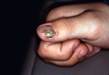
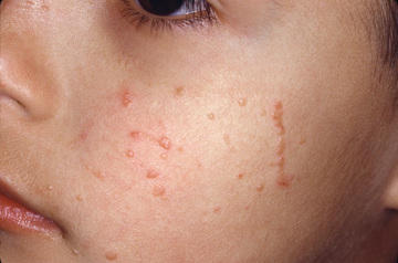
Cutaneous warts develop in 5–10% of children. Common warts (verruca vulgaris), caused most commonly by HPV types 2 and 4, occur most frequently on the fingers, dorsum of the hands (Fig. 687.1 ), paronychial areas, face, knees, and elbows. They are well-circumscribed papules with an irregular, roughened, keratotic surface. When the surface is pared away, many black dots representing thrombosed dermal capillary loops are often visible. Periungual warts are often painful and may spread beneath the nail plate, separating it from the nail bed (Fig. 687.2 ). Plantar warts (verruca plantaris), although similar to the common wart, are caused by HPV type 1 and are usually flush with the surface of the sole because of the constant pressure from weight bearing. When plantar warts become hyperkeratotic (Fig. 687.3 ), they may be painful. Similar lesions (palmar–verruca palmaris) can also occur on the palms. They are sharply demarcated, often with a ring of thick callus. The surface keratotic material must sometimes be removed before the boundaries of the wart can be appreciated. Several contiguous warts (HPV type 4) may fuse to form a large plaque, the so-called mosaic wart. Flat warts (verruca plana), caused by HPV types 3 and 10, are slightly elevated, minimally hyperkeratotic papules that usually remain <3 mm in diameter and vary in color from pink to brown. They may occur in profusion on the face, arms, dorsum of the hands, and knees. The distribution of several lesions along a line of cutaneous trauma (Koebnerization) is a helpful diagnostic feature (Fig. 687.4 ). Lesions may be disseminated in the beard area and on the legs by shaving and from the hairline onto the scalp by combing the hair. Epidermodysplasia verruciformis (EVER1, EVER2 genes), caused primarily by HPV types 5 and 8 (β-papillomaviruses, species 1), manifests as many diffuse verrucous papules. Wart types 9, 12, 14, 15, 17, 25, 36, 38, 47, and 50 may also be involved. Inheritance is thought to be primarily autosomal recessive, but an X-linked recessive form also has been postulated. Warts progress to squamous cell carcinoma in 10% of patients with epidermodysplasia verruciformis.


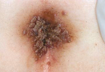
Genital HPV infection occurs in sexually active adolescents, most commonly as a result of infection with HPV types 6 and 11. Condylomata acuminata (mucous membrane warts) are moist, fleshy, papillomatous lesions that occur on the perianal mucosa (Fig. 687.5 ), labia, vaginal introitus, and perineal raphe, and on the shaft, corona, and glans penis. Occasionally, they obstruct the urethral meatus or the vaginal introitus. Because they are located in intertriginous areas, they may become moist and friable. When untreated, condylomata proliferate and become confluent, at times forming large cauliflower-like masses. Lesions can also occur on the lips, gingivae, tongue, and conjunctivae. Genital warts in children may occur after inoculation during birth through an infected birth canal, as a consequence of sexual abuse, or from incidental spread from cutaneous warts. A significant proportion of genital warts in children contain HPV types that are usually isolated from cutaneous warts. HPV infection of the cervix is a major risk factor for the development of carcinoma, particularly if the infection is caused by HPV type 16, 18, 31, 33, 35, 39, 45, 52, 59, 67, 68, or 70. Immunization against types 6, 11, 16, and 18 is available (see Chapter. 417.2 ). Laryngeal (respiratory) papillomas contain the same HPV types as in anogenital papillomas. Transmission is believed to occur from mothers with a genital HPV infection to neonates who aspirate infectious virus during birth and may develop laryngeal papillomatosis.
Histology
The various types of warts share the basic changes of hyperplasia of the epidermal cells and vacuolation of the spinous keratinocytes, which may contain basophilic intranuclear inclusions (viral particles). Warts are confined to the epidermis and do not have “roots.” Individuals with impaired cell-mediated immunity are particularly susceptible to HPV infection. Antibodies occur in response to infection but appear to have little protective effect.
Differential Diagnosis
Common warts are most often confused with molluscum contagiosum. Plantar and palmar warts may be difficult to distinguish from punctate keratoses, corns, and calluses. In contrast to calluses, warts obliterate normal skin markings. Juvenile flat warts mimic lichen planus, lichen nitidus, angiofibromas, syringomas, milia, and acne. Condylomata acuminata may resemble condylomata lata of secondary syphilis.
Treatment
Various therapeutic measures are effective in the treatment of warts. More than 65% of warts disappear spontaneously within 2 yr. Warts are epidermal lesions and do not produce scarring unless they are managed surgically or treated in an overly aggressive fashion. Hyperkeratotic lesions (common, plantar, and palmar warts) are more responsive to therapy if the excess keratotic debris is gently pared with a scalpel until thrombosed capillaries are apparent; further paring induces bleeding. Treatment is most successful when performed regularly and frequently (every 2-4 wk).
Common warts can be destroyed by applications of liquid nitrogen or by pulsed dye laser . Daily application of salicylic acid in flexible collodion or as a stick is a slow but painless method of removal that is effective in some patients. Plantar and palmar warts may be treated with 40% salicylic acid plasters. These should be applied for 5 days at a time with a 2-day rest period between applications. Following removal of the plaster and prolonged soaking in hot water, keratotic debris can be removed with an emery board or pumice stone. Condylomata respond best to weekly applications of 25% podophyllin in tincture of benzoin. The medication should be left on the warts for 4-6 hr and then removed by bathing. Keratinized warts near the genitalia (buttocks) do not respond to podophyllin. Imiquimod (5% cream) applied 3 times weekly is also beneficial. Imiquimod is not only indicated for genital warts, but also has been used successfully to treat warts in other locations; however, it can cause inflammation and irritation, particularly in occluded areas. For nongenital warts, imiquimod should be applied daily. Cimetidine 30-40 mg/kg/day by mouth has been used in children with multiple warts unresponsive to other treatments. Immunotherapy with intralesional candida or Trichophyton antigen may also be employed especially when lesions are numerous or resistant to other therapies. Immunotherapy is performed in clinic and multiple treatments every month (at least 3-4) are usually required. With all types of therapy, care should be taken to protect the surrounding normal skin from irritation. Other treatments include 5-fluorouracil, a chemotherapy agent, which can be helpful, particularly when used with occlusion and sometimes in combination with salicylic acid.
Molluscum Contagiosum
Etiology
The poxvirus that causes molluscum contagiosum is a large double-stranded DNA virus that replicates in the cytoplasm of host epithelial cells. The 3 types cannot be differentiated on the basis of clinical appearance, location of lesions, or a patient's age or sex. Type 1 virus causes most infections. The disease is acquired by direct contact with an infected person or from fomites and is spread by autoinoculation. Children aged 2-6 yr who are otherwise well, and individuals who are immunosuppressed, are most commonly affected. The incubation period is estimated to be 2 wk or longer.
Clinical Manifestations
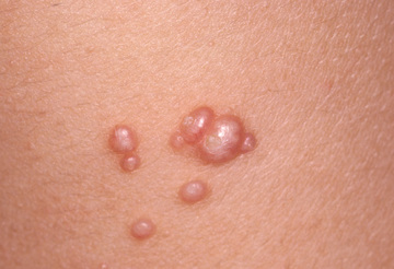
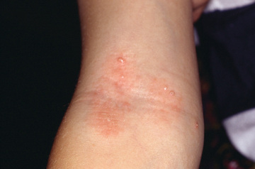
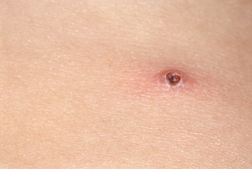
Discrete, pearly, skin-colored, smooth, dome-shaped, papules vary in size from 1 to 5 mm. They typically have a central umbilication from which a plug of cheesy material can be expressed. The papules may occur anywhere on the body, but the face, eyelids, neck, axillae, and thighs are sites of predilection (Fig. 687.6 ). They may be found in clusters on the genitals or in the groin of adolescents and may be associated with other venereal diseases in sexually active individuals. Lesions commonly involve the genital area in children but are not acquired by sexual transmission in most cases. Mild surrounding erythema or an eczematous dermatitis may accompany the papules (Fig. 687.7 ). Lesions on patients with AIDS tend to be large and numerous, particularly on the face. Exuberant lesions may also be found in children with leukemia and other immunodeficiencies. Children with atopic dermatitis are susceptible to widespread involvement in areas of dermatitis. A pustular eruption at the site of individual molluscum lesions is seen (Fig. 687.8 ). It is not a secondary bacterial infection, but rather an immunologic reaction to the molluscum virus, and it should not be treated with antibiotics. Atrophic scars are often seen after this type of reaction.
Differential Diagnosis
Differential diagnosis of molluscum contagiosum includes trichoepithelioma, basal cell carcinoma, ectopic sebaceous glands, syringoma, hidrocystoma, keratoacanthoma, juvenile xanthogranulomas, and warty dyskeratoma. In individuals with AIDS, cryptococcosis may be indistinguishable clinically from molluscum contagiosum. Rarely, coccidioidomycosis, histoplasmosis, or Penicillium marneffei infection masquerades as molluscum-like lesions in an immunocompromised host.
Histology
The epidermis is hyperplastic and hypertrophied, extending into the underlying dermis and projecting above the skin surface. The central plug of material, which is composed of virus-laden cells, may be shelled out from a lesion and examined under the microscope. The rounded, cup-shaped mass of homogeneous cells, often with identifiable lobules, is diagnostic. A specific antibody against molluscum contagiosum virus is detectable in most infected individuals, but it is of uncertain immunologic significance. Cell-mediated immunity is thought to be important in host defense.
Treatment
Molluscum contagiosum is a self-limited disease. The average attack lasts 6-9 mo. However, lesions can persist for years, can spread to distant sites, and may be transmitted to others. Affected patients should be advised to avoid shared baths and towels until the infection is clear. Infection may spread rapidly and produce hundreds of lesions in children with atopic dermatitis or immunodeficiency. Immunotherapy with either candida or trichophyton antigen is the most commonly used treatment . This is repeated every 4 wk until resolution. If lesions are limited in number, then depending on the age of the patient, individual lesions can be treated with liquid nitrogen cryotherapy. For younger children, cantharidin may be applied to the lesions and covered with adhesive bandages to prevent unwanted spread of the blistering agent. A blister forms at the site of application, and the molluscum is removed with the blister. Cantharidin should not be used on the face. Cantharidin availability is very limited or completely unavailable in the United States. Imiquimod has not been proven more effective than placebo in randomized trials. Molluscum is an epidermal disease and should not be overtreated such that scarring results.