CHAPTER 16
Endodontic Management of Primary Teeth
Monty S. Duggal and Hani Nazzal
Pulpal complications such as pain, infection, and loss of teeth at a young age can pose considerable effects to the child’s psychological development [1], quality of life [1,2], growth [3], school attendance, arch space loss [4] in addition to posing considerable morbidity risk [5]. Successful endodontic management of these teeth is therefore crucial in order to maintain these teeth until physiologic exfoliation.
Endodontic management of primary teeth should be considered as part of the overall patient’s management plan. Careful planning including consideration of short‐ as well as long‐term prognosis must be done before any endodontic treatment is considered. Furthermore, other factors such as the effect of the treatment on the underlying permanent tooth germ, patient’s oral hygiene, patient’s medical history and patient’s behavior and future attitudes towards dentistry should be considered before embarking on any endodontic management.
The successful management of pulpally affected primary teeth demands an accurate diagnosis and knowledge of the appropriate endodontic management techniques. Appropriate medical and dental history, radiographic evaluation, and clinical assessment are, therefore, crucial prior to any endodontic management.
Diagnosis of pulpal status of primary teeth
Endodontic management of primary teeth depends on accurate diagnosis of the pulp status; therefore; it is important to differentiate between vital teeth showing signs of reversible pulpitis and those teeth with irreversible pulpitis and necrotic pulps (Box 16.1). Eighty percent of primary teeth with carious exposures but no clinical or radiographic pathology showed inflammation limited to the coronal part of the pulp (chronic coronal pulpitis) [6] (Figure 16.1). Teeth with total chronic pulpitis may show clinical as well as radiographic symptoms, and may not be expected to heal.

Figure 16.1 Histologic view of partial chronic pulpitis of a primary molar. Note the restricted area of chronic inflammation at the site of carious exposure.
Diagnosis of the pulpal status of teeth depends on careful clinical and radiographic assessment of these teeth. Careful assessment of the causative factors, signs, symptoms, and radiographic findings is, therefore; important in order to assess the tooth pulpal status (Box 16.1). This is particularly important where a deep carious lesion is present with breakdown of the marginal ridge.
An understanding of the pulp response to caries as well as correctly interpreting a patient’s symptoms is crucial in deciding whether an indirect pulp capping could be performed or whether more radical intervention would be required. For example, in cases where no pulp exposure is found clinically on removal of all caries for a deep proximal cavity, but where symptoms suggest pulp inflammation, it would be prudent to give consideration to a pulpotomy before restoration of the tooth. However, if there are no symptoms or other signs suggesting pulp inflammation, an indirect pulp capping approach should suffice.
Should endodontic interventions be carried out in the primary dentition?
There are several factors that should be considered when answering this question, such as:
- The pulpal diagnosis
- Occlusal considerations (Box 16.2)
- The patient’s ability to cooperate. Can the treatment be performed conventionally or is sedation/general anesthesia needed?
- Patient’s general and oral health
- Patient’s and parents’ motivation and consent
- Patient’s caries risk
- The risk of injury/infection of the underlying permanent tooth germ
- The effect of the proposed endodontic intervention on the patient’s health, for example infective endocarditis in children who have congenital heart defects or have had heart surgery
- The effect of the proposed endodontic intervention on the underlying permanent tooth germ.
These factors should help the clinician decide whether retaining or extracting the tooth is in the patient’s best interest. The placement of a temporary restoration or stepwise excavation could be considered to monitor the tooth prior to making a decision on endodontic intervention.
Endodontic techniques in primary teeth
The various endodontic techniques are presented in (Box 16.3). All endodontic treatment should be performed with due consideration for pain control and disinfection. Therefore, the tooth should be adequately anesthetized and isolated with a rubber dam.
Since most pulpal treatment of primary teeth involves a vital pulp, it is of utmost importance that the surgical technique is gentle—atraumatic—in order not to decrease the healing capacity of the residual pulp. The recommended technique includes use of diamond burs on high‐speed equipment and irrigation with water in order not to burn the residual pulp. This procedure has been shown under experimental as well as clinical conditions to be the most gentle to the residual pulp [7,8].
The wound treatment is also important. The presence of an extrapulpal blood clot has been shown to interfere with healing and cause chronic inflammation and internal dentin resorption [7]. The wound surface should therefore be gently irrigated with sterile saline in order to achieve hemostasis before application of the medicament. With the increasing use of ferric sulfate as a pulpotomy medicament, achieving hemostasis is no longer a major problem. However, if persistent bleeding is observed even after the application of ferric sulfate, the diagnosis of the state of pulp inflammation should be reconsidered. After pulpal treatment, it is important to provide a tight coronal seal to the oral cavity to prevent leakage and thereby reinfection and future complications.
Stepwise excavation and indirect pulp capping
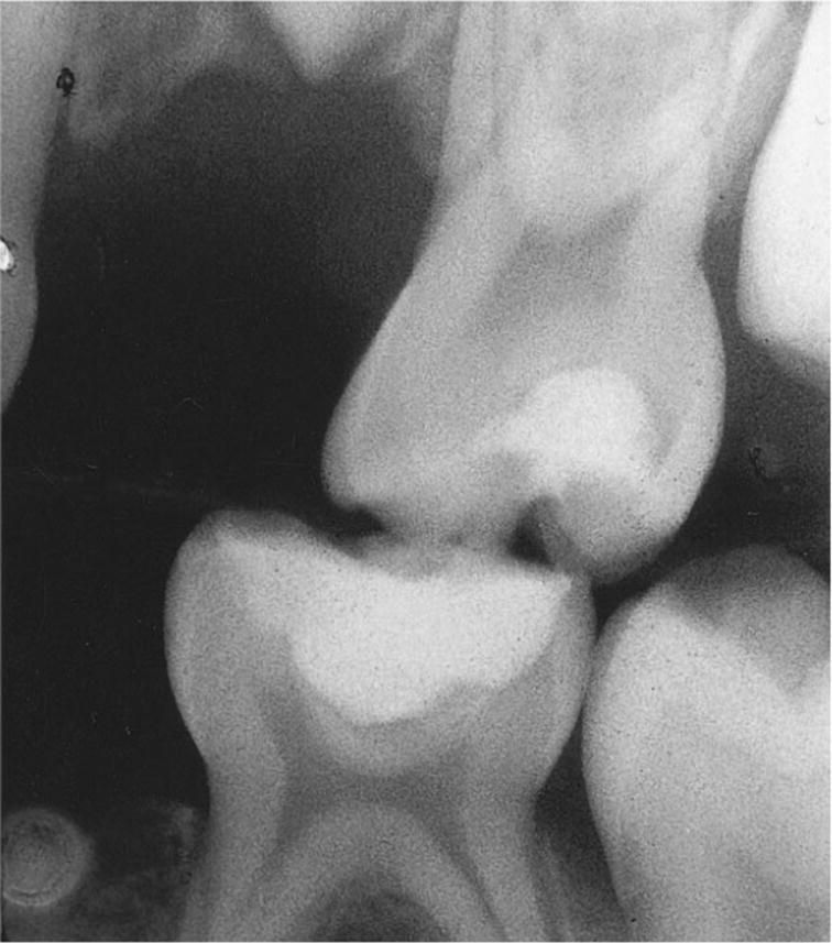
In general, a nonexposed pulp has a more favorable healing capacity than an exposed pulp. However, it has been shown recently that pulpal inflammation in response to proximal caries is more severe as compared to occlusal caries of the same depth (Figure 16.2). This should be an important consideration when planning stepwise excavation or indirect pulp capping for occlusal caries as compared to a deep proximal lesion.

Figure 16.2 Radiograph showing stepwise excavation performed in two primary second molars in order to keep them in situ and without subjective symptoms at least until the permanent first molars are in occlusion.
In the authors’ opinion a more radical approach is often required for a deep proximal lesion as compared to one in an occlusal surface. This is important to consider when treating extensive carious lesions, where an exposure of the pulp may be expected if all carious dentin is removed. If this is expected in proximal caries then a pulpotomy would probably be the preferred option, whereas for occlusal caries, in the same situation, a more conservative approach could be adopted in the absence of any patient symptoms suggestive of pulpitis. Severe caries in first primary molars might lead to poor prognosis for the tooth, and for this particular tooth, extraction should be considered as an alternative to stepwise excavation or any pulp treatment.
The stepwise excavation technique includes removal of most of the soft carious dentin, application of a glass ionomer liner, and sealing the cavity with a semi‐permanent material such as a resin‐reinforced zinc oxide–eugenol cement or glass ionomer cement. The tooth is left for at least 3–6 months, while secondary dentin formation continues, which means less or no risk of exposing the pulp when re‐entering the cavity for final removal of carious dentin and restoration of the tooth [9].
Although stepwise excavation is included in this chapter for the sake of completeness, it is not recommended as a favored approach for the treatment of deep caries in primary molars. The technique requires a second appointment with further administration of local anesthetic to the child. In such cases, if an assessment of pulp inflammation indicates that the patient has reversible pulp inflammation then a one‐visit indirect pulp capping should be considered, with an excellent coronal seal such as a stainless‐steel or an esthetic crown such as zirconia crowns that are widely available these days.
Direct pulp capping
Direct pulp capping is a technique that involves covering the exposed healthy pulp with a medicament, preferably calcium hydroxide, without any surgical intervention. Direct capping should not be considered in primary molars where removal of all caries has resulted in a pulp exposure. The reason is that it has been shown that inflammation of the pulp in primary molars precedes the exposure of the pulp [10,11]. Due to the wide dentinal tubules in primary molars, bacteria penetrate the pulp before it is clinically exposed, causing inflammation. By the time the pulp is clinically exposed, inflammation is usually too extensive for healing to occur with pulp capping, and an inflamed pulp needs to be removed before restoring the tooth. The only situation where direct pulp capping could be considered in primary teeth is where the pulp exposure is traumatic and not due to caries.
Partial pulpotomy
Partial pulpotomy is the treatment of choice only for primary molars with healthy pulps or partial chronic pulpitis (Figure 16.3). However, the diagnosis can be difficult to establish. Partial pulpotomy should be considered in very limited situations where the operator is certain that pulp inflammation is localized to the area just below the site of the exposure. This can be judged by taking a thorough history to assure absence of any signs of pulpitis. Clinically normal bleeding should be observed after the removal of the inflamed pulp from just below the exposure site, and this bleeding should be easily controlled by application of gentle pressure with a cotton pledget. In case more extensive and difficult‐to‐control bleeding is observed, a full coronal pulpotomy should be performed.

Figure 16.3 Radiograph of primary second molar after partial pulpotomy. The hard tissue barrier indicates wound healing; note absence of periradicular pathology.
The technique includes removal of a small part of the pulp just at the site of exposure using high speed and a diamond bur during irrigation with water in order to avoid traumatizing the pulp. Until relatively recently, application of calcium hydroxide on the wound was preferred, but high failure rates have been observed with this material. Nowadays, mineral oxide aggregate (MTA) is considered to be a more biological material for use on the pulp, and many clinicians now prefer its use. The tooth is restored to establish a good coronal seal.
Pulpotomy
Pulpotomy, or vital amputation of the coronal pulp, includes removal of all of the coronal pulp (Box 16.4). The surgical technique and the treatment of the wound are the same as described for partial pulpotomy, but the wound surface is placed at the orifices of the root canals.
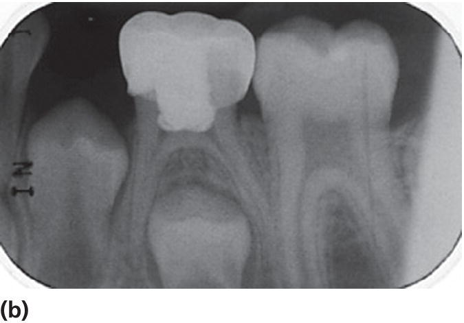
In the authors’ opinion pulpotomy is generally the most accepted option for most pulpally involved primary teeth. There is overwhelming evidence in favor of this technique in the literature over many decades (Figure 16.4).


Figure 16.4 Pulpotomy performed in lower left second primary molar with pulp exposure and pulpitis (a). The tooth was maintained in the arch with healthy bone in the bifurcation (b), after it was restored with a stainless steel crown (c).
Pulpectomy and root canal therapy
A pulpectomy should be considered if the pulp is considered to be irreversibly inflamed in the whole tooth including the root canals, necrotic pulp or in presence of infection. Although all pediatric dentists should have this technique in their repertoire, its use is extremely limited. One situation is where the second premolar is missing, while retention of the primary tooth is indicated due to orthodontic considerations. In many situations clinicians choose to avoid pulpectomy and root canal therapy in the primary dentition and to choose extraction.
The number of root canals for primary molars is generally the same as those for permanent molars except that there is an increased prevalence of two distal canals in lower molars, especially the lower second primary molar.
The pulp chamber is accessed in the same way as for a pulpotomy, root canals identified and gently cleaned with endodontic files. Hand‐held files are used, with rotary root canal instruments never used in primary molars. Various medicaments have been advocated for filling in the root canals, such as pure zinc oxide–eugenol or a mixture of calcium hydroxide and iodoform. The most important property of any material used in root canals of primary teeth is that it should fully resorb with physiologic resorption of the roots of the primary tooth.
As mentioned (Box 16.2) the risk for occlusal complications is rare and should be seen in relation to the risk for the underlying permanent tooth germ if perforating the roots and/or pushing infected material through the apical foramen.
Wound dressings and tissue reactions
A proper wound dressing for use in pediatric endodontics should not induce any persistent pathologic changes of the residual pulp or the periapical tissues. In the case of primary teeth the wound dressing should not carry any risk to the underlying tooth germ. Two main groups of wound dressings have for a long time been the choice: calcium hydroxide and formocresol. For some years now ferric sulfate has increasingly been used and more recently mineral trioxide aggregate (MTA) has been introduced (Box 16.5) [12].
Calcium hydroxide preparations were used because of their biological effect, i.e., stimulation of a hard tissue barrier, which allows healing in an organ such as the pulp, which lacks epithelium. For the pulp treatment in the primary teeth poor outcomes have been reported when calcium hydroxide has been used and most clinicians would seldom consider its use for pulp therapy. MTA is a new wound dressing material with promising results.
The use of formocresol has been questioned for several years. Since the International Agency for Research on Cancer [13] classified formaldehyde as carcinogenic to humans, efforts have been reinforced to prevent formocresol being used in endodontic treatment. The present trend is away from devitalizing medicaments and towards searching for medicaments able to keep primary teeth with total chronic pulpitis or partial necrosis in the dentition without causing any periapical pathologic changes. Ferric sulfate belongs to this category and has so far showed similar success rates as formocresol. In this context, the possibility of extraction of primary teeth with more advanced pathologic pulpal changes should be seriously considered unless there are specific indications where the preservation of the primary tooth in the arch is deemed essential.
Calcium hydroxide and MTA
Calcium hydroxide is strongly alkaline with a pH about 12 and initially causes a chemical injury to the vital pulp with a zone of firm necrosis adjacent to vital pulp tissue. The response of the residual pulp tissue is characteristic of wounded connective tissue. It begins with a vascular and inflammatory reaction to control and eliminate the irritating agent. Thereafter, the repair process starts, including proliferation of cells and formation of new collagen. When the pulp is protected from irritation, new odontoblasts differentiate and the newly formed tissue assumes a dentin‐like appearance, which indicates that pulp function has been restored. Mineralization of the newly formed collagen starts with dystrophic calcification of the zone with firm necrosis leading to deposition of mineral in the newly formed collagen as well [7].
MTA induces similar tissue reactions when applied to pulp tissue. MTA, when mixed with water, forms crystals of calcium oxide in amorphous structure of 33% Ca2+, 49% PO43−, 2% C, 3% Cl−, and 6% Si2+. MTA was developed as an apex filling material, but has also been proven successful in vital pulp therapy procedures [14]. It is a powder that sets in the presence of moisture and has a pH of 12.5, similar to calcium hydroxide. It has an ability to actively promote hard tissue formation.
The hard tissue barrier is a criterion of wound healing and must be considered favorable because it facilitates the clinical handling of the tooth. The hard tissue barrier does not mean a tight seal against the oral cavity. It is therefore of utmost importance that the restoration of the tooth provides a tight seal to prevent infection through microleakage.
Calcium hydroxide and MTA contribute to wound healing but, as with any other pulpal medicament, they have no healing effect on chronically inflamed pulp tissue. Therefore; the diagnosis of the residual pulp and the surgical technique are of paramount importance, even more so than the medicament used. In primary teeth, medicaments such as MTA must be restricted to teeth where the residual pulp is considered to be healthy in order to achieve a favorable outcome.
Formocresol
The use of formocresol as pulp medicament should be avoided because of its negative systemic properties. Formaldehyde has a known carcinogenic, immunogenic, toxic, and mutagenic potential which makes it questionable and unsuitable for use in pedodontic endodontics [13,15]. Formaldehyde is the devitalizing ingredient in formocresol. Formaldehyde, the aqueous solution of the gas formaldehyde, is used for fixation of tissue in histologic studies. The same process is considered to occur in vivo. The clinical procedure includes a pulpotomy, followed by application of cotton pellets moistened with formocresol to the root pulps at the canal openings for 5 minutes. The treated root pulps and the furcation area are then covered with slow‐setting zinc oxide–eugenol cement in order to get a bacteria‐tight seal and the tooth is restored.
The rationale for use of this type of wound dressing is to create a chemically altered zone at the medicament interface that will be stable over time and leave the deeper untreated pulp tissue vital and uninflamed. Studies show, however, that the degree of penetration of formaldehyde is time and dose dependent [16]. Furthermore, clinical–histologic investigations have shown that chronic inflammation or even partial necrosis of the residual pulp often results [17].
In conclusion, formocresol does not induce healing of the pulp but does cause pathologic changes in the residual pulp. Formocresol very seldom gives rise to clinical complications but in light of various concerns that have been expressed in the literature, it is extremely difficult to justify its use in children.
Ferric sulfate
Ferric sulfate (15.5% Fe2SO4) has been used as pulpotomy agent as a substitute for formocresol for 15–20 years. Ferric sulfate in contact with blood forms a ferric ion–protein complex, which seals the cut blood vessels mechanically, producing hemostasis [18]. The effect of ferric sulfate is hemostatic but not bactericidal or fixative. After application of ferric sulfate for 15 seconds, the pulp is covered with zinc oxide–eugenol and the cavity sealed. Research has shown similar results both regarding clinical, radiographic, and histologic results as formocresol. Healing has not been achieved, but the teeth could be retained in the dentition for shorter or longer intervals. There are no known systemic risks of using ferric sulfate in pulpal treatment.
Complications (primary teeth)
Internal dentin resorption is a common complication irrespective of medicament used, but is often reported to be caused by calcium hydroxide. However, investigations have shown that the reasons for resorption are most probably chronic inflammation of the residual pulp and/or the presence of an extrapulpal blood clot on the wound surface before capping with calcium hydroxide [7].
MTA has not been used in the primary dentition for a long enough time to allow estimation of complications with any certainty.
Ferric sulfate has no fixative effect on the pulp and is merely a hemostatic solution. Therefore, an accurate diagnosis of the residual pulp after removal of what is considered as inflamed is essential. If the pulp left behind after a partial or a coronal pulpotomy is in a state of chronic inflammation then internal resorption could manifest in the months following pulp therapy.
Follow‐up and long‐term prognosis
Long‐term prognosis might be an inappropriate term considering primary teeth. It is, however, reasonable to consider the necessity for and outcome of the treatment planned. It is not ethically justifiable to let a child undergo treatment, perhaps experienced as both difficult and stressful, if the outcome is uncertain or poor.
Stepwise excavation should seldom be considered as it means multiple episodes of treatment for the same tooth for the child patient. There is little evidence to support the use of this technique in the primary dentition. Partial pulpotomy should be considered in cases with the symptomless cariously exposed pulp. Partial pulpotomy does include some strain on the young patient, but the treatment is usually short, since the surgical intervention is minimal, and control of bleeding is easy because no large vessels are cut. Compared to direct capping, the possibility to seal the cavity efficiently is better and consequently the risk of future bacterial contamination and failure is less. According to the literature, the prognosis for partial pulpotomy of primary molars is about 80% after 1 year [19] and 75% after 2 years, which is acceptable.
Pulpotomy (coronal pulpotomy), using ferric sulfate or MTA, is the treatment of choice for most cases where pulpal treatment is required. Success rates (no clinical or radiographic signs of loss of vitality) of over 85% over 2 years have been reported with the use of ferric sulfate and over 95% when MTA is used [19].
Handling the emergency child patient
In this chapter, discussion of emergency treatment will be restricted to the preschool child with toothache due to extensive carious lesions. For treatment of acute trauma and the use of sedation and pain control the reader is referred to Chapters 9 and 18.
Symptoms may vary depending on the extent of the carious lesion and the involvement of the pulp. The aim of the emergency treatment is to provide pain control and to eliminate the cause of pain. The pain control usually includes local analgesia and may also include sedation and oral analgesics. History taking should focus on the general health of the child and how and when the pain is occurring, followed by the examination of the mouth and the teeth.
After the diagnosis has been made, a decision is made for conservative treatment or extraction. Should the final treatment be performed immediately or should temporary measures be taken? Is sedation necessary? Are antibiotics necessary? Finally, the parents must be given thorough information before treatment to be able to give an informed consent.
Pain at food intake
The tooth is painful when the child is eating, especially sour, hot, or sweet food. In such a case the tooth usually has an extensive carious lesion with retentive capacity. This is why the pain appears when ingredients of the food irritate the pulp when stuck in the cavity. The radiograph shows a deep carious cavity, not necessarily with pulp lesion.
The emergency treatment involves stepwise excavation avoiding exposure of the pulp, application of calcium hydroxide, and a temporary filling. Ledermix, which is a combination of antibiotics and a steroid, has also been suggested as an alternative to calcium hydroxide, but the use of Ledermix is controversial in certain countries. However, its use in emergency situations is recommended in the UK [20]. The parents (guardian) should be provided with some analgesics and information about caries‐preventive measures; later the tooth is restored with a more permanent filling material.
Toothache at night
This type of pain is usually not related to food intake but appears spontaneously, especially at night, and the child wakes up. Symptoms commonly have been apparent for a period of time and the child has been avoiding drinking and eating on the painful side of the mouth and might, therefore, be tired and difficult to handle. The tooth is mostly grossly carious, has some distinct clinical symptoms such as tenderness on percussion and/or pressure and increased mobility, indicating involvement of the periodontal membrane. Spontaneous pain that is not relieved by analgesics is usually an indication of advanced pulp pathology. Although X‐rays may not show pathologic changes, widening of the periodontal space is usually seen, indicating more advanced pathologic changes. The treatment may include some temporary measures but the final treatment is extraction of the primary tooth. When there are definite radiographic signs of periradicular and/or interradicular periodontitis, the tooth is extracted with due consideration to sedation and pain control (see case description in Box 16.6).
The emergency patient with toothache may also show general symptoms such as fever and tiredness. The oral symptoms are often more pronounced, with involvement of the regional lymph nodes and swelling of the oral tissues surrounding the tooth. Evaluation of the nature of the swelling includes the size and extent of the swelling, and the location, intraoral and/or extraoral. Is the swelling increasing? There are distinct pathologic radiographic signs and purulence might be present. The child may be in need of antibiotics. Temporary measures may include opening the tooth, exposing the pulp, and draining the pus. The tooth should not be left open to the oral environment, risking further infection, but should be sealed temporarily and may have to be extracted as soon as it is possible to do so.
The rationale for being radical in the emergency situation is based on the value of the tooth and the consideration that the child should not suffer more strain than necessary. If an extraction is performed with adequate sedation, preferably with amnesic effect, and analgesia, the emergency treatment is adequate and the child will soon recover. There is very seldom any need for several treatment sessions of pulp treatment with doubtful outcome as these usually end up with extraction anyway. Concern about the child’s future attitude to dental care is more important than the child losing a painful tooth.
Use of antibiotics
Antibiotics should always be used restrictively, which is also the case in children’s dentistry. There are medical conditions that demand antibiotic cover even for ordinary dental treatment and the reader is referred to Chapters 23 and 24 regarding medically compromised children. With emergency treatment, however, there are situations where antibiotics may be indicated, most often for the child’s general state or when a tooth is seriously infected with a risk of spread to neighboring tissues, teeth, tooth germs, and ultimately a risk of a general spreading to more vital organs.
In general, penicillin V (phenoxymethylpenicillin) or amoxicillin is the drug of choice, but with anaerobic microorganisms, which is often the case in oral infection, metronidazole may be used. Metronidazole is to be preferred when there is an infected pulp, for instance following a luxation injury. Although the treatment with calcium hydroxide may be bacteriostatic, there is a risk of persistent infection of periradicular tissues.
Erythromycin is the third alternative with dental infections in children. It is used in cases where there is an allergy to penicillin V. In some countries clindamycin is recommended as an alternative to erythromycin.
Dosage may vary between different countries. Therefore clinicians are advised to check drug doses and regimens concerning children, contraindications, and drug interactions according to national guidelines prior to prescribing any medication.
References
- 1. Casamassimo P, Thikkurissy S, Edelstein B, Maiorini E. Beyond the dmft: the human and economic cost of early childhood caries. J Am Dent Assoc 2009;140:650–7.
- 2. Clinical Affairs Committee—Pulp Therapy Subcommittee: Guideline on Pulp Therapy for Primary and Immature Permanent Teeth: 2014. http://www.aapd.org/media/Policies_Guidelines/G_Pulp.pdf (accessed 18.5.2015).
- 3. Clarke M, Locker D, Berall G, Pencharz P, Kenny D, Judd P. Malnourishment in a population of young children with severe early childhood caries. Pediatr Dent 2006;28:254–9.
- 4. Lin Y, Lin W, Lin Y. Immediate and six‐month space changes after premature loss of a primary maxillary first molar. J Am Dent Assoc 2007;138:362–8.
- 5. Levine R, Pitts N, Nugent Z. The fate of 1587 unrestored carious deciduous teeth: a retrospective general dental practice based study from Northern England. Br Dent J 2002;193:99–103.
- 6. Schröder U. Agreement between clinical and histologic findings in chronic coronal pulpitis in primary teeth. Scand J Dent Res 1977;85:583–7.
- 7. Schröder U. Effects of calcium hydroxide‐containing pulpcapping agents on pulp cell migration, proliferation and differentiation. J Dent Res 1985;64:541–8.
- 8. Schröder U, Szpringer‐Nodzak M, Janicha J, Wacinska M, Budny J, Mlosek K. A one‐year follow‐up of partial pulpotomy and calcium hydroxide capping in primary molars. Endod Dent Traumatol 1987;3:304–6.
- 9. Bjorndal L. Indirect pulp therapy and stepwise excavation. J Endod 2008;34:S29–33.
- 10. Duggal M. Response of the primary pulp to inflammation: a review of the Leeds studies and challenges for the future. Eur J Paediatr Dent 2002;3:111–14.
- 11. Desponia K, Day P, High A, Duggal M. Histological comparison of pulpal inflammation in primary teeth with occlusal or proximal caries. Int J Paediatr Dent 2008;19:23–6.
- 12. Smail‐Faugeron V, Courson F, Durieux P, Muller‐Bolla M, Glenny AM, Fron Chabouis H. Pulp treatment for extensive decay in primary teeth. Cochrane Database Systematic Reviews 2014: http://onlinelibrary.wiley.com/doi/10.1002/14651858.CD003220.pub2/abstract (accessed 18.5.2015).
- 13. International Agency for Research on Cancer: Press Release No. 153: June 15, 2004. http://www.iarc.fr/en/media‐centre/pr/2004/pr153.html (accessed 18.5.2015).
- 14. Qudeimat M, Barrieshi‐Nusair K, Owais A. Calcium Hydroxide Vs. Mineral trioxide aggregates for partial pulpotomy of permanent molars with deep caries. Eur Arch Paediatr Dent 2007;8:99–104.
- 15. Lewis B. Formaldehyde in dentistry: a review for the millennium. J Clin Pediatr Dent 1998;22:167–77.
- 16. Mejàre I, Hasselgren G, Hammarström L. Effect of formaldehyde‐containing drugs on human dental pulp evaluated by enzyme histochemical technique. Scand J Dent Res 1976;84:29–36.
- 17. Rölling I, Thylstrup A. A 3‐year clinical follow‐up study of pulpotomized primary molars treated with the formocresol technique. Scand J Dent Res 1975;83:47–57.
- 18. Fischer D. Tissue Management. A new solution to an old problem. Gen Dent 1987;35:178–82.
- 19. Huth K, Paschos E, Hajek‐Al‐Khatar N, Hollweck R, Crispin A, Hickel R, et al. Effectiveness of 4 pulpotomy techniques–randomized controlled trial. J Dent Res 2005 12:1144–8.
- 20. Rodd HD, Waterhouse PJ, Fuks AB, Fayle SA, Moffat MA. Pulp therapy for primary molars. Int J Paediatr Dent 2006;16 Suppl 1:15–23.
- 21. Markovic D ZV, Vucetic M. Evaluation of three pulpotomy medicaments in primary teeth. Eur J Paediatr Dent 2005 6:133–8.