Fig. 28.1 Arteries of the upper limb
Right limb, anterior view.
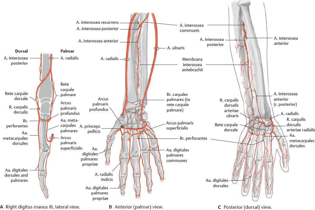
Fig. 28.4 Arteries of the forearm and hand
Right limb. The aa. ulnaris and radialis are interconnected by the arcus palmares superficialis and profundus, the rr. perforantes, and the rete carpale dorsale.
Fig. 28.7 Fossa cubitalis
Right limb, anterior view. The subcutaneous veins of the fossa cubitalis have a highly variable course.
Venipuncture
The veins of the fossa cubitalis are frequently used when drawing blood. In preparation, a tourniquet is applied above the fossa cubitalis. This allows arterial blood to flow, but blocks the return of venous blood. The resulting swelling makes the veins more visible and palpable.
 Lymph from the upper limb and mamma drains to the nll. axillares. The superficial lymphatics of the upper limb lie in the subcutaneous tissue, while the deep lymphatics accompany the arteries and deep veins. Numerous anastomoses exist between the two systems.
Fig. 28.9 Lymphatic drainage of the hand
Right hand, radial view. Most of the hand drains to the nll. axillares via nll. cubitales. However, the pollex, index, and dorsum manus drain directly.
Fig. 28.10 Nodi lymphoidei axillares
Right side, anterior view. For surgical purposes, the nll. axillares are divided into three levels with respect to their relationship with the m. pectoralis minor: lateral (level I), posterior (level II), or medial (level III). They have major clinical importance in breast cancer (see p. 77).
Almost all muscles in the upper limb are innervated by the plexus brachialis, which arises from segmenta C5–T1 medullae spinalis. The rr. anteriores of the nn. spinales give off direct branches (pars supraclavicularis of the plexus brachialis) and merge to form three trunci, six divisiones (three anteriores and three posteriores), and three fasciculi. The pars infraclavicularis of the plexus brachialis consists of short branches that arise directly from the fasciculi and long (terminal) branches that traverse the limb.

The supraclavicular branches of the plexus brachialis arise directly from the radices plexus brachialis (rr. anteriores of the nn. spinales) or from the trunci plexus brachialis in the regio cervicalis lateralis.
Table 28.2 Pars supraclavicularis plexus brachialis
Nerve |
Level |
Innervated muscle |
N. dorsalis scapulae |
C4–C5 |
M. levator scapulae Mm. rhomboidei major et minor |
N. suprascapularis |
C4–C6 |
M. supraspinatus M. infraspinatus |
N. subclavius |
C5–C6 |
M. subclavius |
N. thoracicus longus |
C5–C7 |
M. serratus anterior |
The fasciculus posterior gives off three short branches (arising at the level of the fasciculi plexus brachialis) and two long branches (terminal nerves, see pp. 368–369).
Table 28.3 Branches of the fasciculus posterior
Nerve |
Level |
Innervated muscle |
Short branches |
||
Upper n. subscapularis |
C5–C6 |
M. subscapularis |
Lower n. subscapularis |
M. subscapularis M. teres major |
|
N. thoracodorsalis |
C6–C8 |
M. latissimus dorsi |
Long (terminal) branches |
||
N. axillaris |
C5–C6 |
See p. 368 |
N. radialis |
C5–T1 |
See p. 369 |
The n. axillaris may be damaged in a fracture of the collum chirurgicum of the humerus. This results in limited ability to abduct the arm and may cause a loss of profile of the shoulder.
Table 28.4 Nervus axillaris (C5–C6)
Motor branches |
Innervated muscles |
Rr. musculares |
M. deltoideus M. teres minor |
Sensory branch |
|
N. cutaneus brachii lateralis superior |
|
Table 28.5 Nervus radialis (C5–T1)
Motor branches |
Innervated muscles |
Rr. musculares |
M. brachialis (partial) |
M. triceps brachii |
|
M. anconeus |
|
M. brachioradialis |
|
Mm. extensores carpi radialis longus and brevis |
|
R. profundus (terminal br.: n. interosseus antebrachii posterior) |
M. supinator |
M. extensor digitorum |
|
M. extensor digiti minimi |
|
M. extensor carpi ulnaris |
|
Mm. extensores pollicis brevis and longus |
|
M. extensor indicis |
|
M. abductor pollicis longus |
|
Sensory branches |
|
Rr. articulares from n. radialis: Capsula articularis of the art. humeri |
|
Rr. articulares from n. interosseus antebrachii posterior: Capsula articularis of the carpus and four radial artt. metacarpophalangeae |
|
N. cutaneus brachii posterior |
|
N. cutaneus brachii lateralis inferior |
|
N. cutaneus antebrachii posterior |
|
Rr. superficiales |
Nn. digitales dorsales |
R. communicans ulnaris |
|
Chronic n. radialis compression in the axilla (e.g., due to extended/improper crutch use) may cause loss of sensation or motor function in the hand, forearm, and posterior arm. More distal injuries (e.g., during anesthesia) affect fewer muscles, potentially resulting in wrist drop with intact m. triceps brachii function.
The fasciculi medialis and lateralis plexus brachialis give off four short branches. The nn. intercostobrachiales are included with the short branches of the plexus brachialis, although they are actually the cutaneous branches of the nn. intercostales 2 and 3.
Table 28.7 Nervus musculocutaneus (C5–C7)
Motor branches |
Innervated muscles |
Rr. musculares |
M. coracobrachialis |
M. biceps brachii |
|
M. brachialis |
|
Sensory branches |
|
N. cutaneus antebrachii lateralis |
|
Rr. articulares: Capsula articularis of the art. cubiti (anterior part) |
|
Note: N. musculocutaneus innervation of the arm is purely motor; innervation of the forearm is purely sensory. |
|
The n. medianus is a terminal branch arising from both the fasciculi medialis and lateralis plexus brachialis. The n. ulnaris arises exclusively from the fasciculus medialis.
N. medianus injury caused by fracture/dislocation of the art. cubiti may result in compromised grasping ability and sensory loss in the fingertips (see Fig. 28.23 for territories). See also carpal tunnel syndrome (p. 387).
Table 28.8 Nervus medianus (C6–T1)
Motor branches |
Innervated muscles |
Direct rr. musculares |
M. pronator teres |
M. radialis flexor carpi |
|
M. palmaris longus |
|
M. flexor digitorum superficialis |
|
Rr. musculares from n. interosseus antebrachii anterior |
M. pronator quadratus |
M. flexor pollicis longus |
|
M. flexor digitorum profundus (radial half) |
|
Thenar r. muscularis |
M. brevis abductor pollicis |
Caput superficiale musculi flexoris pollicis brevis |
|
M. opponens pollicis |
|
Rr. musculares from nn. digitales palmares communes |
Mm. lumbricales I and II |
Sensory branches |
|
Rr. articulares: Capsulae articulares of the artt. cubiti and carpi |
|
R. palmaris of n. medianus (eminentia thenaris) |
|
R. communicans cum nervo ulnari |
|
Nn. digitales palmares communes |
|
Nn. digitales palmares proprii |
|
Table 28.9 Nervus ulnaris (C7–T1)
Motor branches |
Innervated muscles |
Direct rr. musculares |
M. flexor carpi ulnaris |
M. flexor digitorum profundus (ulnar half) |
|
R. muscularis from superior n. ulnaris |
M. palmaris brevis |
Rr. musculares from r. profundus |
M. abductor digiti minimi |
M. brevis flexor digiti minimi |
|
M. opponens digiti minimi |
|
Mm. lumbricales III and IV |
|
Mm. interossei palmares and dorsales |
|
M. adductor pollicis |
|
Caput profundum musculi flexoris pollicis brevis |
|
Sensory branches |
|
Rr. articulares: Capsulae articulares of the artt. cubiti, carpi, and metacarpophalangeae |
|
R. dorsalis (terminal brs.: nn. digitales dorsales) |
|
R. palmaris |
|
Nn. digitales palmares proprii (from r. superficialis) |
|
N. digitales palmares communes (from r. superficialis; terminal brs.: nn. digitales palmares proprii) |
|
Ulnar nerve palsy is the most common peripheral nerve damage. The n. ulnaris is most vulnerable to trauma or chronic compression in the art. cubiti and ulnar tunnel (see p. 387). Nerve damage causes “clawing” of the hand and atrophy of the mm. interossei. Sensory losses are often limited to the 5th digit.
Fig. 28.29 Posterior shoulder
Right shoulder, posterior view. Raised: M. trapezius (pars transversa). Windowed: M. supraspinatus. Revealed: Suprascapular region.

Fig. 28.33 Anterior shoulder: Deep dissection
Right limb, anterior view. Removed: Mm. sternocleidomastoideus, omohyoideus, and pectoralis major. This dissection reveals the neurovascular contents of the regio cervicalis lateralis (see p. 642) and axilla (see pp. 380–381).
Table 28.11 Walls of the axilla
Anterior wall |
M. pectoralis major M. pectoralis minor Fascia clavipectoralis |
Lateral wall |
Sulcus intertubercularis of humerus |
Posterior wall |
M. subscapularis M. teres major M. latissimus dorsi |
Medial wall |
Lateral thoracic wall M. serratus anterior |

Fig. 28.35 Regio brachialis
Right arm, anterior view. Removed: Mm. deltoideus, pectorales major et minor. Revealed: Sulcus bicipitalis medialis.
Fig. 28.38 Regio antebrachii posterior
Right forearm, anterior view during pronation. Reflected: Mm. anconeus and triceps brachii. Removed: Mm. extensor carpi ulnaris and extensor digitorum.
Fig. 28.41 Canalis carpi: Cross section
Right hand, proximal view. The tight fit of sensitive neurovascular structures with closely apposed, frequently moving tendons in the canalis carpi often causes problems (carpal tunnel syndrome) when any of the structures swell or degenerate.
Fig. 28.46 Cutaneous innervation of the dorsum of the hand (dorsum manus)
Right hand, posterior view.
Fig. 28.47 Anatomic snuffbox
Right hand, radial view. The three-sided “anatomic snuffbox” is bounded by the tendons of insertion of the m. abductor pollicis longus and mm. extensores pollicis brevis and longus.


 Clinical box 28.1


