32 Knee & Lower Leg
Tibia & Fibula
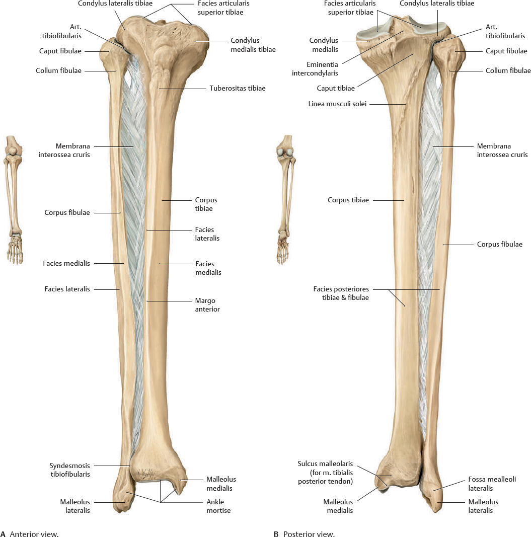
 The tibia and fibula articulate at two joints, allowing limited motion (rotation). The membrana interossea cruris is a sheet of tough connective tissue that serves as an origin for several muscles in the leg. It also acts with the syndesmosis tibiofibularis to stabilize the art. talocruralis.
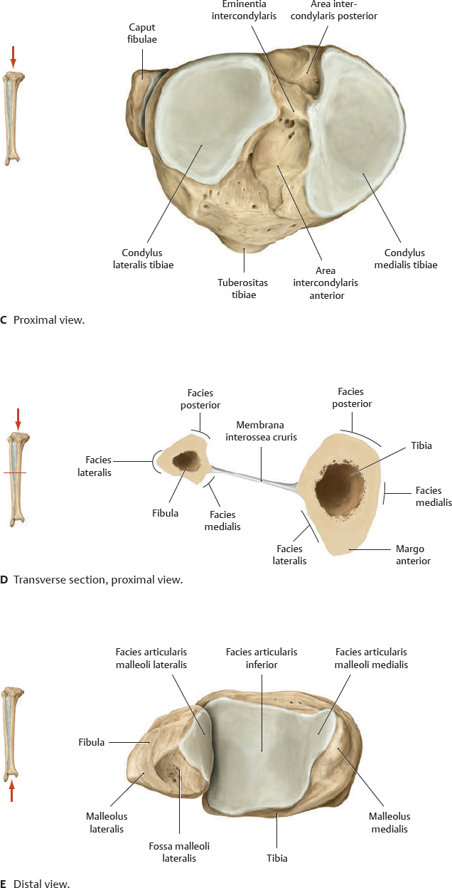
Fig. 32.1 Tibia and fibula
Right lower leg.
 Clinical box 32.1
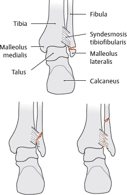
Fibular fracture
When diagnosing a fibular fracture, it is important to determine whether the syndesmosis tibiofibularis (see p. 426) is disrupted. Fibular fractures may occur distal to, level with, or proximal to the syndesmosis tibiofibularis; the latter two frequently involve tearing of the syndesmosis.
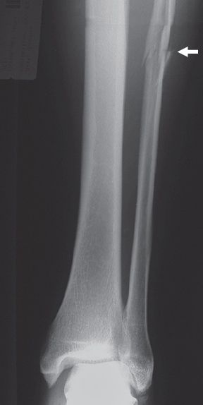
In this fracture located proximal to the syndesmosis (arrow), the syndesmosis is torn, as indicated by the widened medial joint space of the upper art. talocruralis (see pp. 450–451).
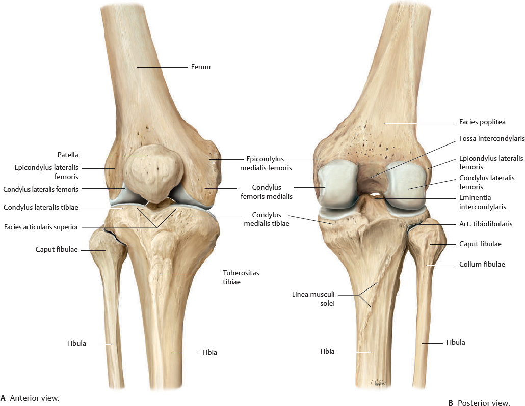
Articulatio Genus: Overview
In the art. genus, the femur articulates with the tibia and patella. Both joints are contained within a common capsula articularis and have communicating cavitates articulares. Note: The fibula is not included in the art. genus (contrast to the radius in the art. cubiti; see p. 322). Instead, it forms a separate rigid articulation with the tibia.
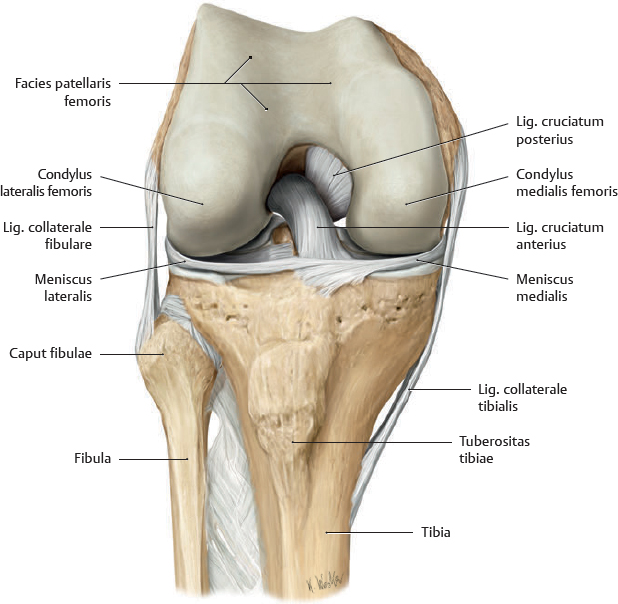
Fig. 32.2 Right articulatio genus
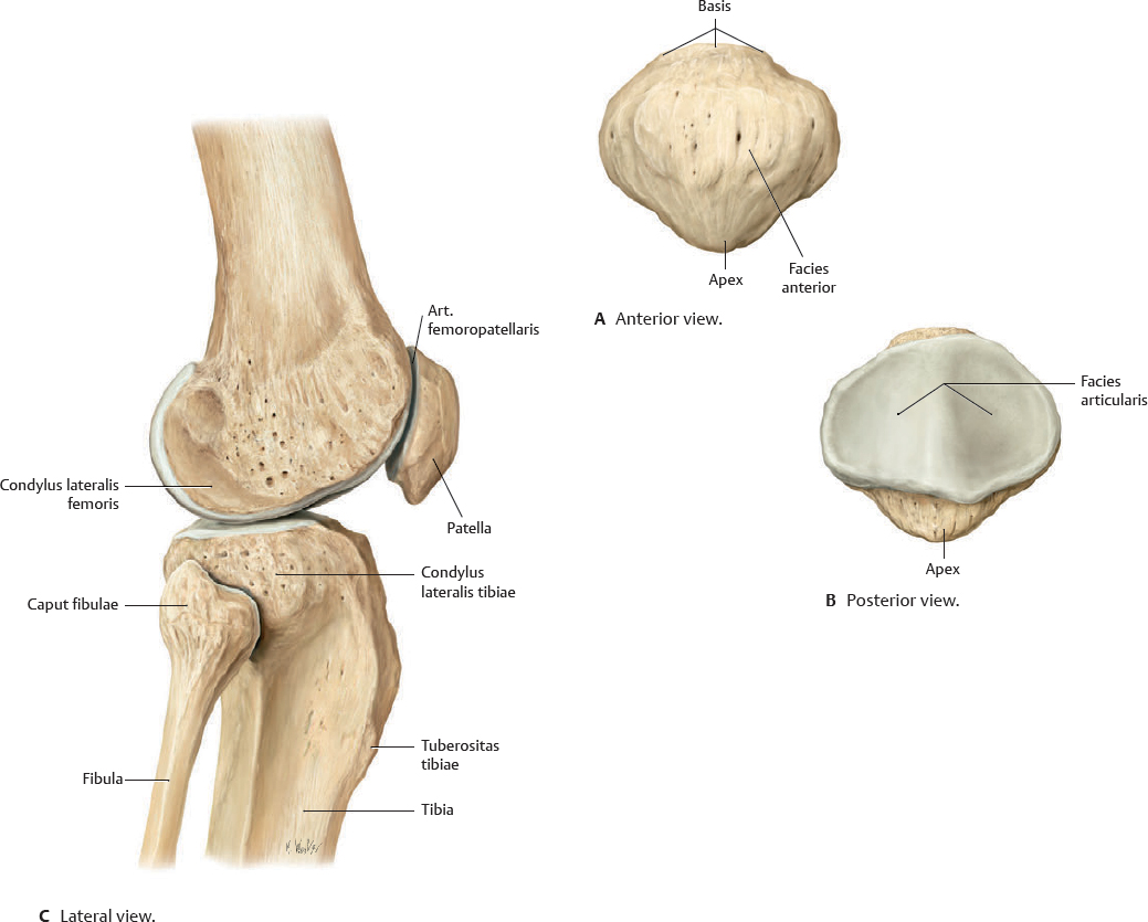
Fig. 32.4 Articulatio femoropatellaris
Transverse section through art. femoropatellaris. Distal view with right knee in slight flexion.
Articulatio Genus: Capsula Articularis, Ligaments & Bursae
Table 32.1 Ligaments of the articulatio genus
Extrinsic ligaments |
Anterior side |
Lig. patellae |
Retinaculum patellae mediale |
Retinaculum patellae laterale |
Retinaculum patellae transversale mediale |
Retinaculum patellae transversale laterale |
Medial and lateral sides |
Lig. collaterale tibiale |
Lig. collaterale fibulare |
Posterior side |
Lig. popliteum obliquum |
Lig. popliteum arcuatum |
Intrinsic ligaments |
Lig. cruciatum anterius |
Lig. cruciatum posterius |
Lig. transversum genus |
Lig. meniscofemorale posterius |
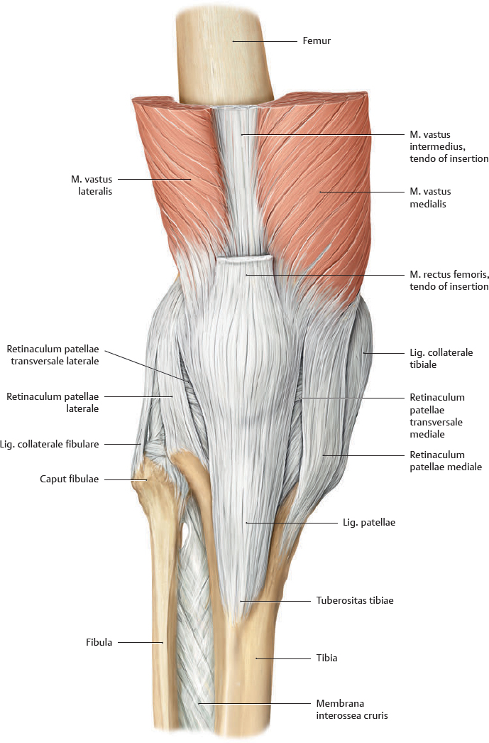
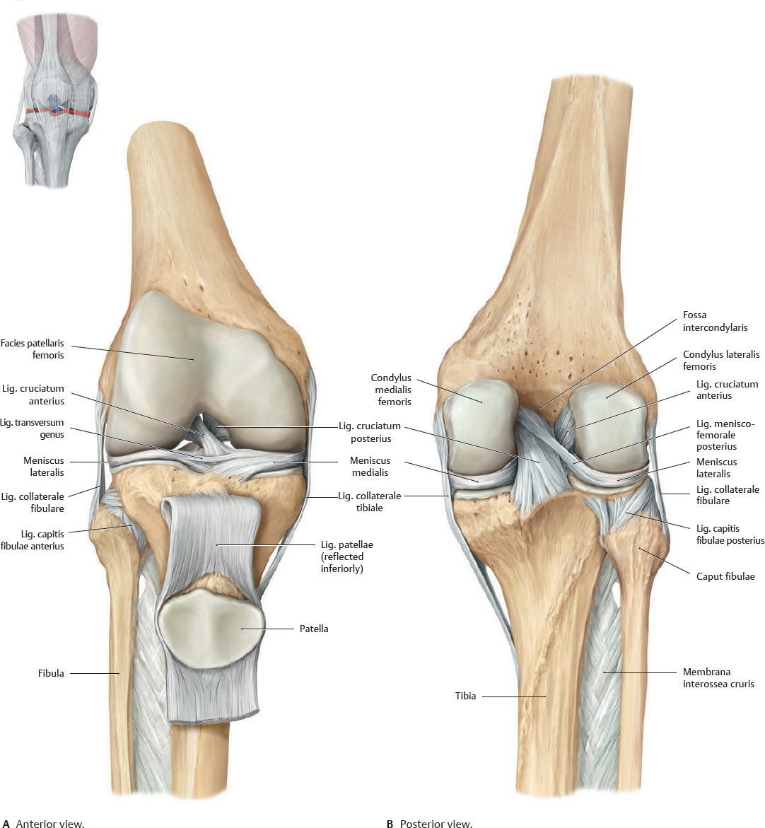
Fig. 32.5 Ligaments of the articulatio genus
Anterior view of right knee.
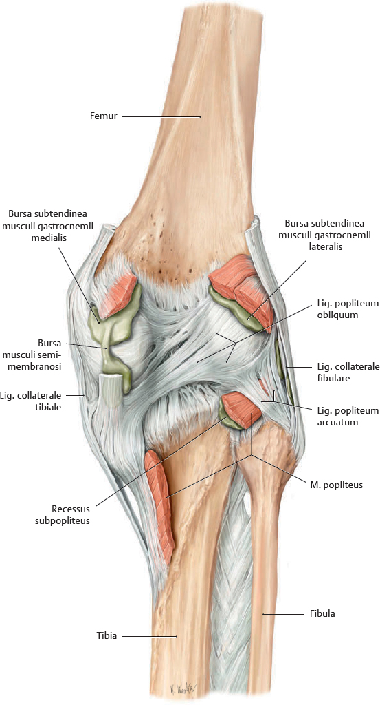
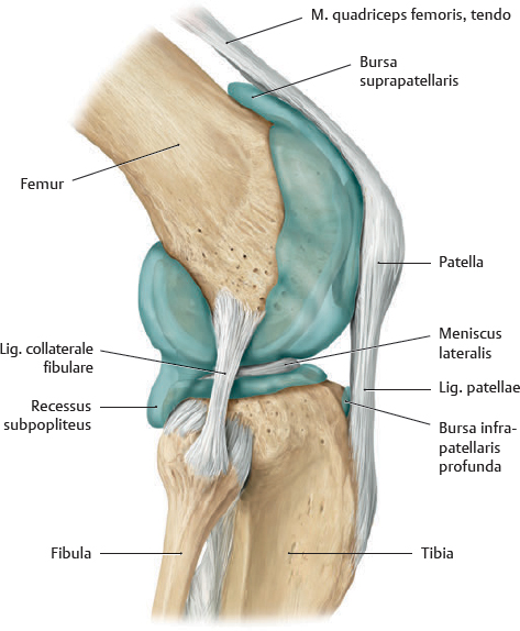
Fig. 32.6 Capsula articularis, ligaments, and periarticular bursae
Posterior view of right knee. The cavitas articularis communicates with peri-articular bursae at the recessus subpopliteus, bursa musculi semimembranosi, and bursa subtendinea musculi gastrocnemii lateralis.
Clinical box 32.2
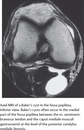
Gastrocnemio-semimembranosus bursa (Baker's cyst)
Painful swelling behind the genu may be caused by a cystic outpouching of the capsula articularis (synovial popliteal cyst). This frequently results from an increase in intra-articular pressure (e.g., in rheumatoid arthritis).
Articulatio Genus: Ligaments & Menisci
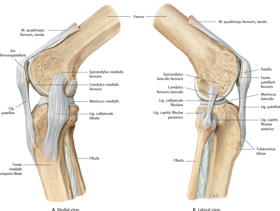
Fig. 32.7 Ligamenta collateralia and patellaria of the articulatio genus
Right art. genus. Each art. genus has ligg. collateralia tibiale and fibulare. The lig. collaterale tibiale is attached to both the capsula articularis and the meniscus medialis, whereas the lig. collaterale fibulare has no direct contact with either the capsula articularis or the meniscus lateralis. Both ligg. collateralia are taut when the knee is in extension and stabilize the joint in the coronal plane.
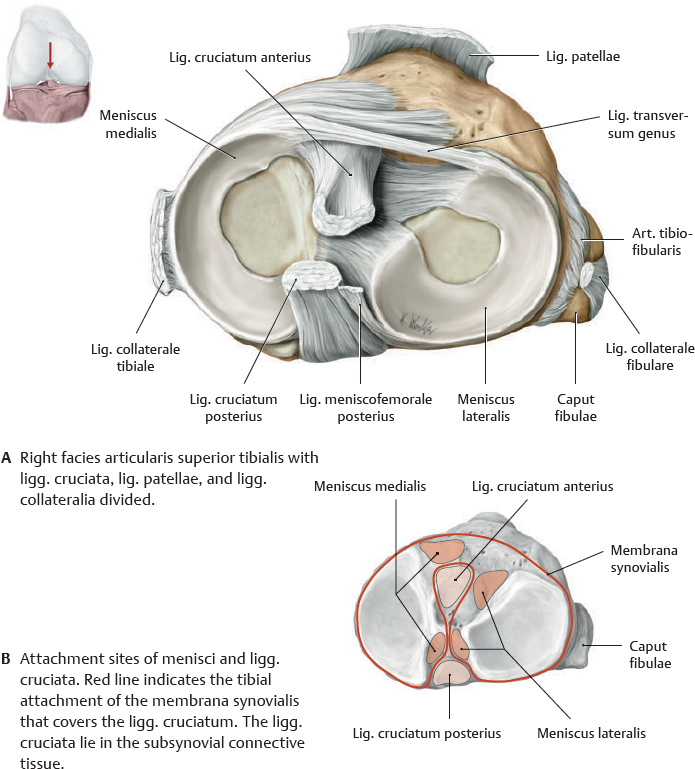
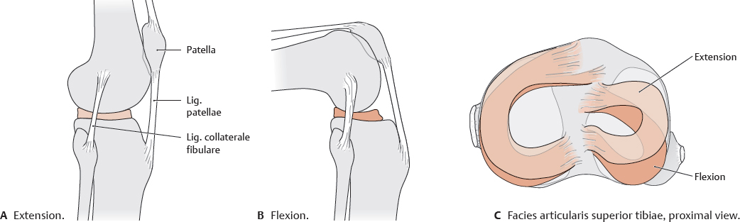
Fig. 32.8 Menisci in the articulatio genus
Right facies articularis superior tibiae, proximal view.
Clinical box 32.3
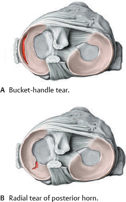
Injury to the menisci
The less mobile meniscus medialis is more susceptible to injury than the meniscus lateralis. Trauma generally results from sudden extension or rotation of the flexed knee while the leg is fixed.
Fig. 32.9 Movements of the menisci
Right art. genus.
Ligamenta Cruciata
Fig. 32.10 Ligamenta cruciata and collateralia
Right art. genus. The ligg. cruciata keep the facies articulares of the femur and tibia in contact, while stabilizing the art. genus primarily in the sagittal plane. Portions of the ligg. cruciata are taut in every joint position.
Fig. 32.11 Right articulatio genus in flexion
Anterior view with capsula articularis and patella removed.
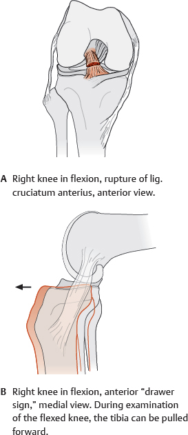
Clinical box 32.4
Rupture of ligamenta cruciata
Lig. cruciatum rupture destabilizes the art. genus, allowing the tibia to move forward (anterior “drawer sign”) or backward (posterior “drawer sign”) relative to the femur. Lig. cruciatum anterius ruptures are approximately 10 times more common than lig. cruciatum posterius ruptures. The most common mechanism of injury is an internal rotation trauma with the leg fixed. A lateral blow to the fully extended knee with the foot planted tends to cause concomitant rupture of the ligg. cruciatum anterius and collaterale fibulare, as well as tearing of the attached meniscus medialis.
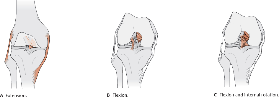
Fig. 32.12 Ligamenta cruciata and collateralia in flexion and extension
Right knee, anterior view. Taut ligament fibers in red.
Cavitas Articularis Articulationis Genus
Fig. 32.13 Cavitas articularis
Right knee, lateral view. The cavitas articularis was demonstrated by injecting liquid plastic into the art. genus and later removing the capsula.
Fig. 32.14 Opened capsula articularis
Right knee, anterior view with patella reflected downward.
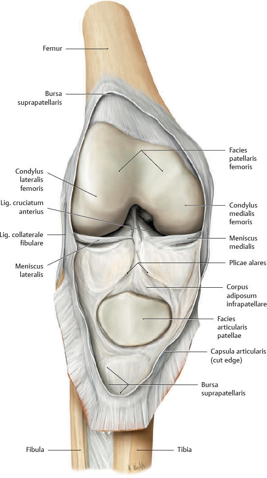
Fig. 32.15 Attachments of the capsula articularis
Right art. genus, anterior view.
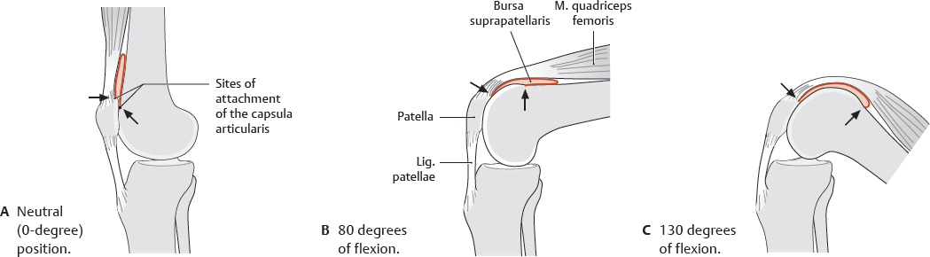
Fig. 32.16 Bursa suprapatellaris during flexion
Right art. genus, medial view.
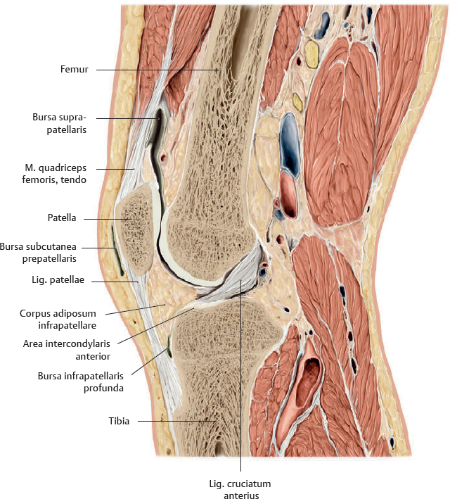
Fig. 32.17 Right articulatio genus: Midsagittal section
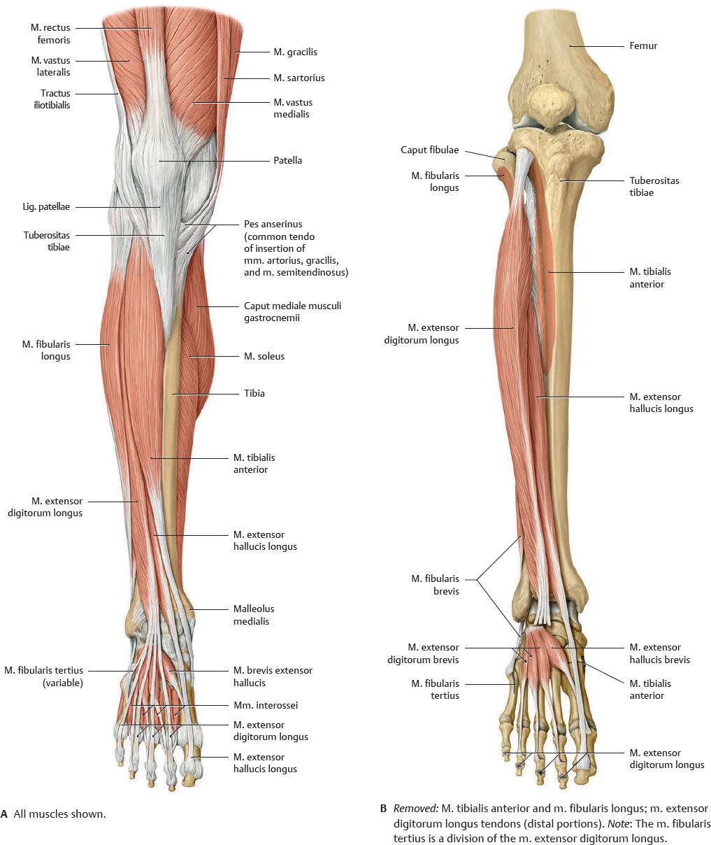
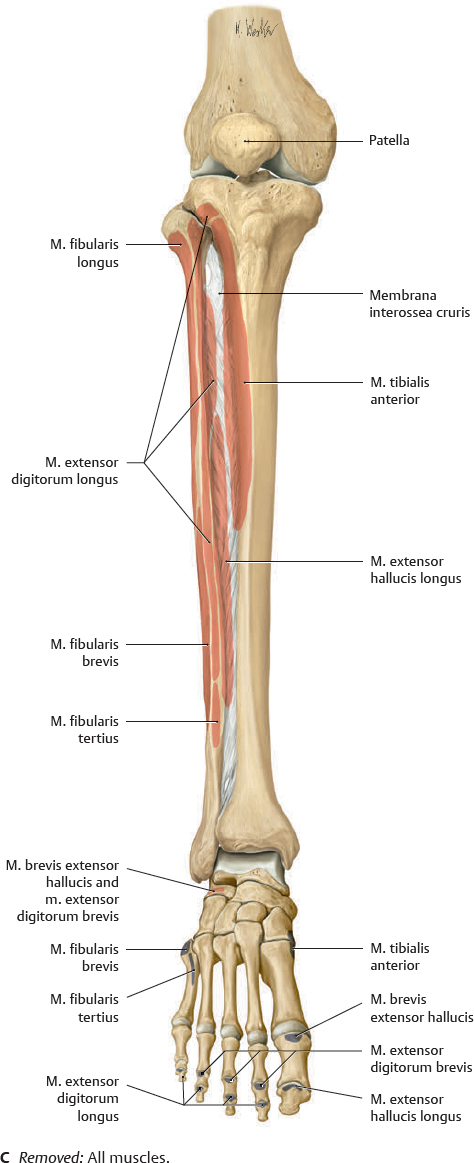
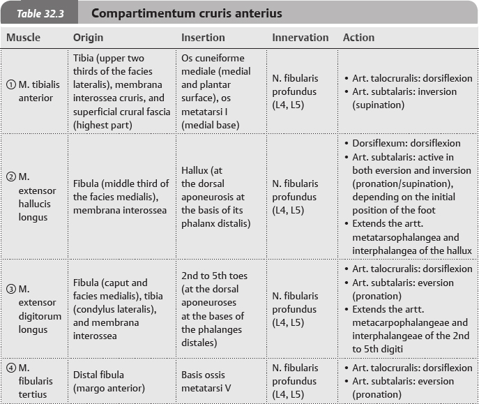
Muscles of the Lower Leg: Compartimenta Anterius et Laterale
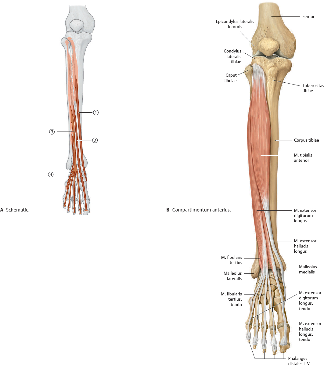
Fig. 32.18 Muscles of the compartimentum cruris anterius
Right lower leg. Muscle origins shown in red, insertions in blue.
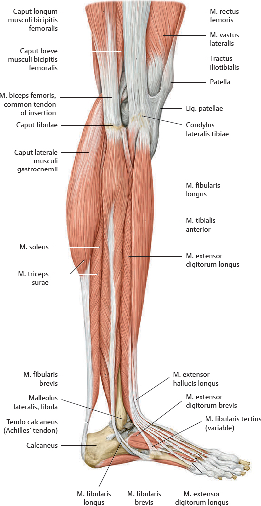
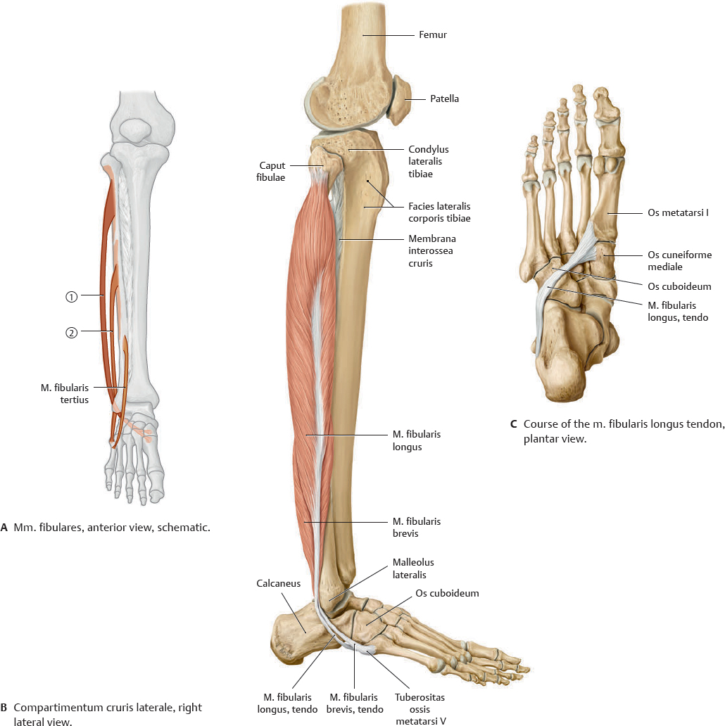
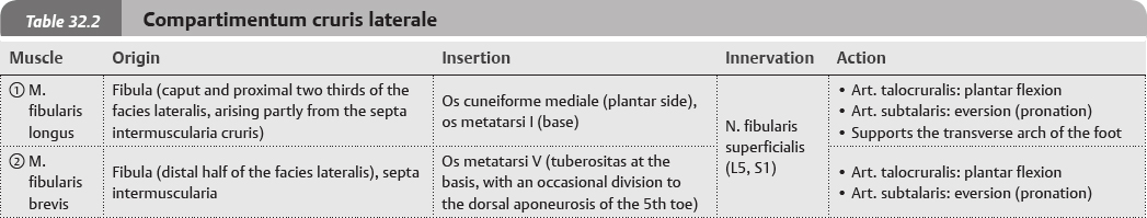
Fig. 32.19 Muscles of the compartimentum cruris laterale
Right lower leg. The m. triceps surae is composed of the m. soleus and two heads of the m. gastrocnemius.
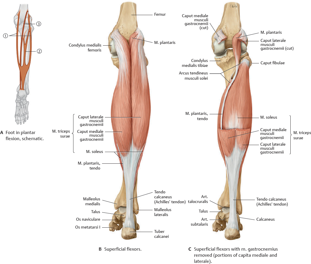
Muscles of the Lower Leg: Compartimentum Posterius
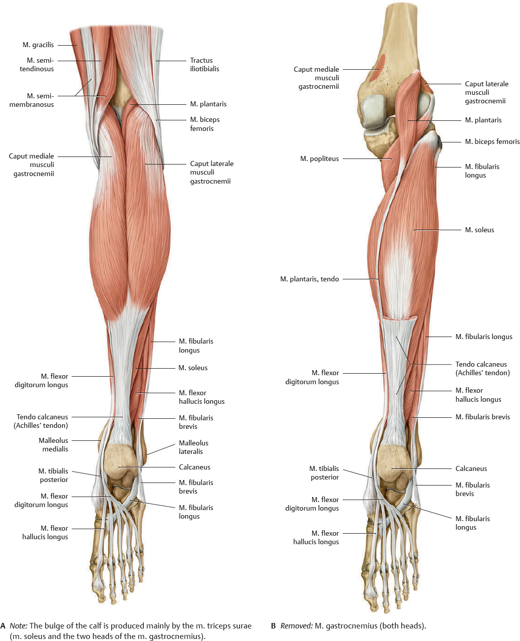
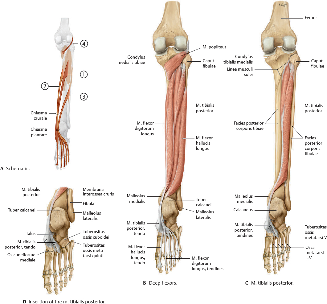
Fig. 32.20 Muscles of the compartimentum cruris posterius
Right lower leg. Muscle origins shown in red, insertions in blue.
Muscle Facts (I)
The muscles of the lower leg control the flexion/extension and inversion/eversion of the foot, which provide stability to the lower limb during movements at the artt. genus and coxae.
Fig. 32.21 Muscles of the compartimentum cruris laterale
Right lower leg and foot.
Fig. 32.22 Muscles of the compartimentum cruris anterius
Right lower leg, anterior view.
Muscle Facts (II)
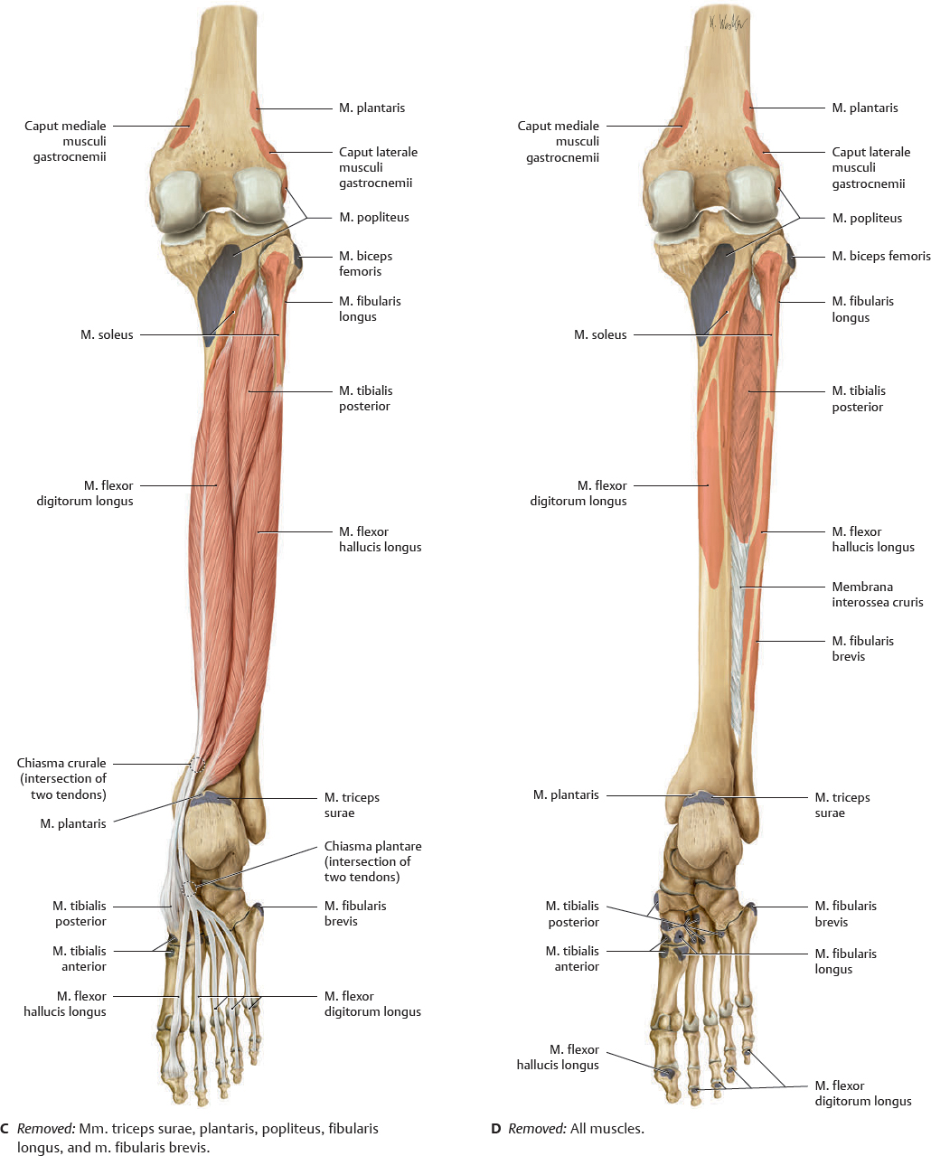
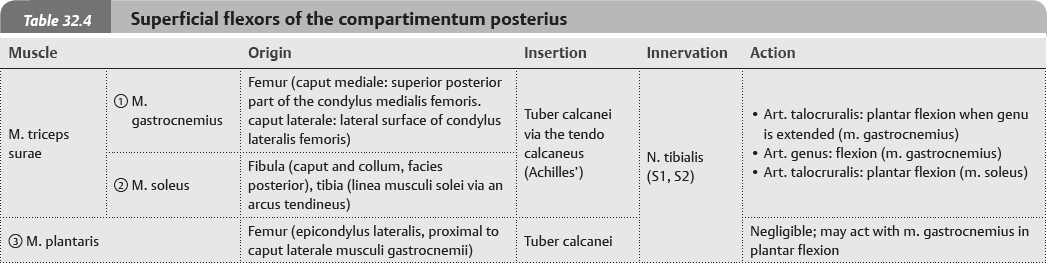
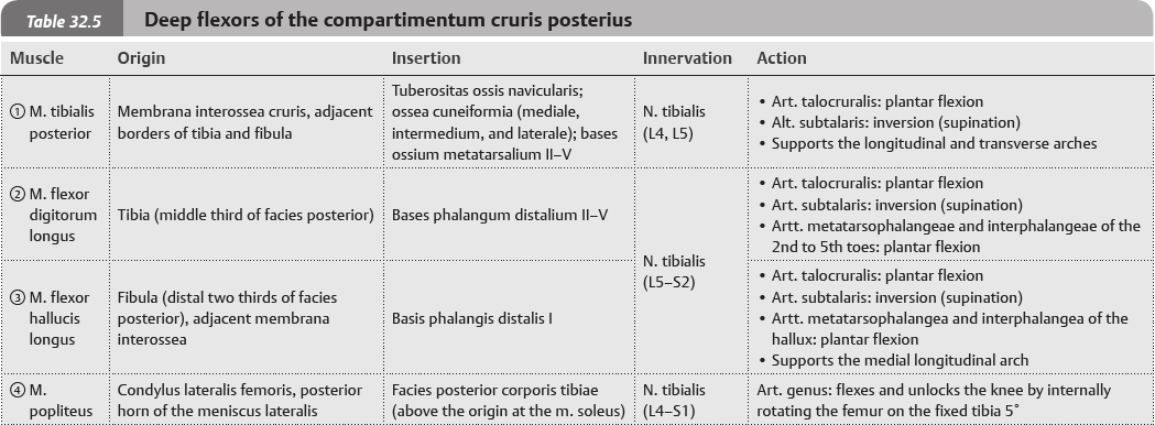
The muscles of the compartimentum posterius are divided into two groups: the superficial and deep flexors. These groups are separated by the septum intermusculare cruris posterius.
Fig. 32.23 Muscles of the compartimentum cruris posterius: Superficial flexors
Right lower leg, posterior view.
Fig. 32.24 Compartimentum cruris posterius: Deep flexors
Right lower leg with foot in plantar flexion, posterior view.