IN CONTEXT
Biology
c.1600 The first compound microscope is developed in the Netherlands, probably by either Hans Lippershey or Hans and Zacharius Janssen.
1644 Italian priest and self-taught scientist Giovanni Battista Odierna produces the first description of living tissue, using a microscope.
1674 Antonie van Leeuwenhoek is the first to see single-celled organisms under the microscope.
1682 Leeuwenhoek observes the nuclei inside the red blood cells of salmon.
1931 The invention of the electron microscope by Hungarian physicist Leó Szilárd allows much higher resolution images to be made.
The development of the compound microscope in the 17th century opened up a whole new world of previously unseen structures. A simple microscope consists of just one lens, while the compound microscope, developed by Dutch spectacle makers, uses two or more lenses, and generally provides greater magnification.
English scientist Robert Hooke was not the first to observe living things using a microscope. However, with the publication of his Micrographia in 1665, he became the first bestselling popular science author, stunning his readers with the new science of microscopy. Accurate copperplate drawings made by Hooke himself showed objects the public had never seen before – the detailed anatomies of lice and fleas; the compound eyes of a fly; the delicate wings of a gnat. He also drew some man-made objects – the sharp point of a needle appeared blunt under the microscope – and used his observations to explain how crystals form and what happens when water freezes. The English diarist Samuel Pepys called Micrographia “the most ingenious book that I ever read in my life”.
Describing cells
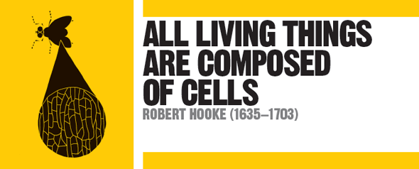
One of Hooke’s drawings was of a thin slice of cork. In the structure of the cork, he noted what looked like the walls dividing monks’ cells in a monastery. These were the first recorded descriptions and drawings of cells, the basic units from which all living things are made.

Hooke’s drawings of dead cork cells show empty spaces between the cell walls – living cells contain protoplasm. He calculated that there were more than a billion cells in 16cm3 (1in3) of cork.
See also: Antonie van Leeuwenhoek • Isaac Newton • Lynn Margulis