IN CONTEXT
Biology
1869 Friedrich Miescher first identifies DNA, in blood cells.
1920s Phoebus Levene and others analyse the components of DNA as sugars, phosphates, and four types of base.
1944 Experiments show DNA to be a carrier of genetic data.
1951 Linus Pauling proposes the alpha helix structure for certain biological molecules.
1963 Frederick Sanger develops sequencing methods to identify bases along DNA.
1960s DNA’s code is cracked: three DNA bases of code for each amino acid in a protein.
2010 Craig Venter and his team implant artificially made DNA into a living bacterium.
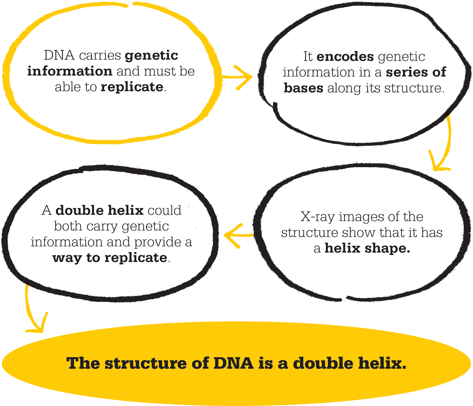
In April 1953, the answer to a fundamental mystery about living organisms appeared in a short article published without fanfare in the scientific journal, Nature. The article explained both how genetic instructions are held inside organisms and how they are passed on to the next generation. Crucially, it described, for the first time, the double-helix structure of deoxyribose nucleic acid (DNA), the molecule that contains the genetic information.
The article was written by James Watson, a 29-year-old American biologist, and his older British research colleague, biophysicist Francis Crick. Since 1951, they had jointly been working on the challenge of DNA’s structure at the Cavendish Laboratory, University of Cambridge, under its director, Sir Lawrence Bragg.
DNA was the hot topic of the day, and an understanding of its structure seemed so tantalizingly within reach that by the early 1950s, teams in Europe, the USA, and the Soviet Union were vying to be the first to “crack” DNA’s three-dimensional shape – the elusive model that allowed DNA simultaneously to carry genetic data in some kind of chemically coded form, and to replicate itself completely and accurately, so that the same genetic data was passed to offspring, or daughter cells, including those of the next generation.
The past in DNA
The DNA molecule was not discovered in 1953, as is often popularly thought, nor were Crick and Watson the first to find out what it was made from. DNA has a much longer history of research. In the 1880s, the German biologist Walther Flemming had reported that “X”-like bodies (later named chromosomes) appeared inside cells as the cells were preparing to divide. In 1900, Gregor Mendel’s experiments with heredity in pea plants were rediscovered – Mendel had been the first to suggest that there were units of heredity that came in pairs (which would later be called genes). At about the same time as Mendel was being rediscovered, breeding experiments by American physician Walter Sutton and, independently, by German biologist Theodor Boveri revealed that sets of chromosomes (the rod-shaped structures that carry genes) pass from a dividing cell to each of its daughter cells. The ensuing Sutton–Boveri theory proposed that chromosomes are the carriers of genetic material.
Soon, more scientists were investigating these mysterious X-shaped bodies. In 1915, American biologist Thomas Hunt Morgan showed that chromosomes were indeed the carriers of hereditary information. The next step was to look at the constituent molecules of chromosomes – molecules that might be candidates for genes.
"So beautiful it has to be true."
James Watson
New pairs of genes
In the 1920s, two types of candidate molecules were discovered: proteins called histones, and nucleic acids, which had been described chemically in 1869 as nuclein by Swiss biologist Friedrich Miescher. The Russian-American biochemist Phoebus Levene and others gradually identified the main ingredients of DNA in increasing detail as nucleotide units, each made up of a deoxyribose sugar, a phosphate, and one of four sub-units called bases. By the end of the 1940s, the basic formula of DNA as a giant polymer – a huge molecule consisting of repeating units, or monomers – was clear. By 1952, experiments with bacteria had shown that DNA itself, and not its rival candidates, the proteins inside chromosomes, was the physical embodiment of genetic information.
Tricky research tools
The competing researchers were using several advanced research tools, including X-ray diffraction crystallography, in which X-rays were passed through a substance’s crystals. A crystal’s unique geometry in terms of its atomic content made the X-ray beams diffract, or bend, as they passed through. The resulting diffraction patterns of spots, lines, and blurs were captured on photographic film. Working backwards from those patterns, it was possible to work out the structural details within the crystal. This was not an easy task. X-ray crystallography has been likened to studying the myriad light patterns cast by a crystal chandelier on the ceiling and walls of a large room, and using them to work out the shapes and positions of each piece of glass in the chandelier.
"It is one of the more striking generalizations of biochemistry…that the twenty amino acids and the four bases, are, with minor reservations, the same throughout Nature."
Francis Crick
Pauling in the lead
The British research team at the Cavendish Laboratory was eager to beat the American researchers, led by Linus Pauling. In 1951, Pauling and his colleagues Robert Corey and Herman Branson had already achieved a breakthrough in molecular biology when they correctly proposed that many biological molecules – including haemoglobin, the oxygen-carrying substance in blood – have a corkscrew-like helix shape. Pauling named this molecular model the alpha helix.
Pauling’s breakthrough had pipped the Cavendish Laboratory at the post, and it looked as though the precise shape of DNA’s structure was within his grasp. Then, early in 1953, Pauling proposed that the structure of DNA was in the form of a triple helix.
By this time, James Watson was working at the Cavendish Laboratory. He was only 25 years old, but he had the enthusiasm of youth and two degrees in zoology, and had studied the genes and nucleic acids of bacteriophages – the viruses that infect bacteria. Crick, aged 37, was a biophysicist with an interest in the brain and neuroscience. He had studied proteins, nucleic acids, and other giant molecules in living things. He had also observed the Cavendish team racing to beat Pauling to the alpha helix idea, and later analysed their mistaken suppositions and dead-end exploratory efforts.
Both Watson and Crick also had experience of X-ray crystallography, albeit in different areas, and together they soon began musing on two questions that fascinated them both: how does DNA as a physical molecule encode genetic information, and how is this information translated into the parts of a living system?
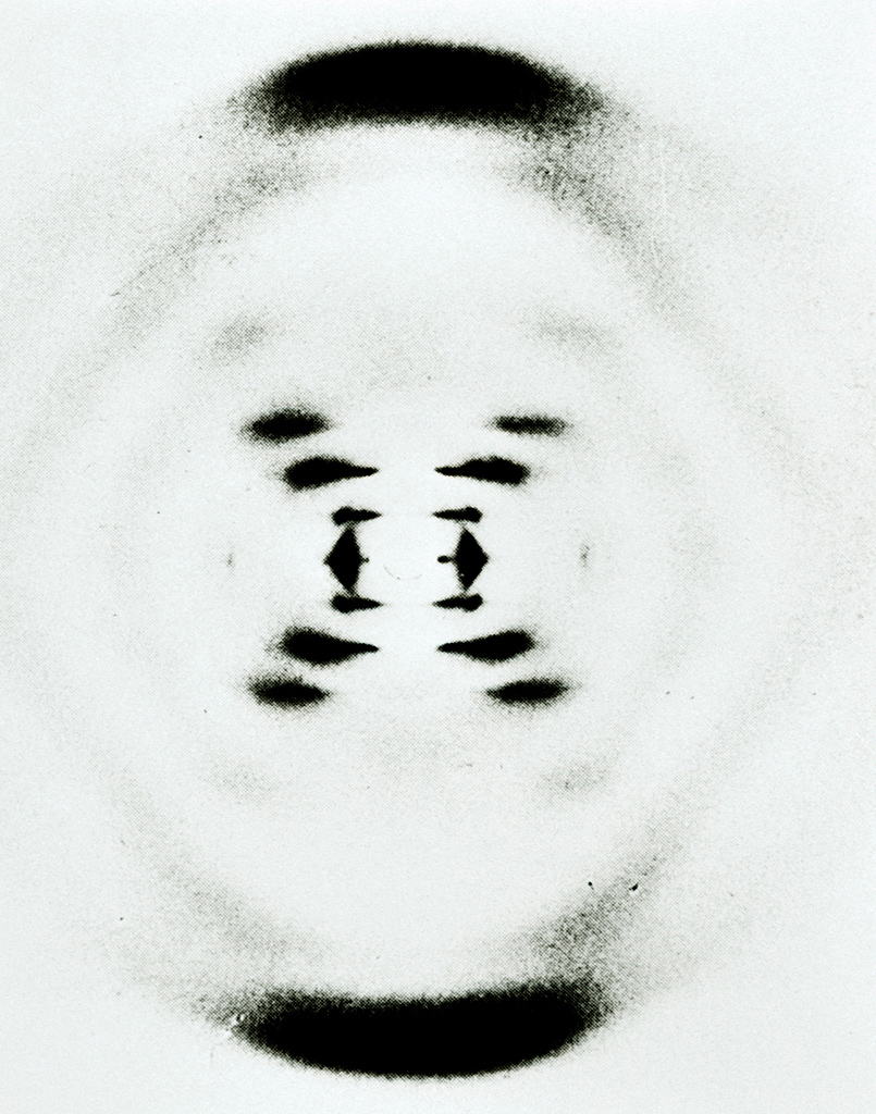
This X-ray diffraction photograph of DNA was obtained by Rosalind Franklin in 1953, and was the biggest clue to cracking DNA. The helical structure of DNA was ascertained from the pattern of spots and bands.
Crucial crystal pictures
Watson and Crick knew of Pauling’s success with the alpha helix model of proteins, in which the molecule twisted along a single corkscrew path, repeating its main structure every 3.6 turns. They also knew that the latest research evidence did not seem to support Pauling’s triple helix model for DNA. This led them to wonder whether the elusive model was one that was neither a single nor a triple helix. The two conducted hardly any experiments themselves. Instead they collected data from others, including the results of chemical experiments that gave information about the angles of the links, or bonds, between the various ingredient atoms and subgroups of DNA. They also pooled their joint knowledge of X-ray crystallography and approached those researchers who had made the highest-quality images of DNA and other similar molecules. One such image was “photo 51”, which became key to their achieving their breakthrough.
Photo 51 was an X-ray diffraction image of DNA that resembled an “X” seen through the slats of a Venetian blind – fuzzy to our eyes, but at that time, among the sharpest and most informative of DNA’s X-ray pictures. Some debate surrounds the identity of the photographer who took this historic picture. It came from the laboratory of a British biophysicist named Rosalind Franklin, an expert in X-ray crystallography, and her graduate student Raymond Gosling, at King’s College, London. Each has been credited with the image at various times.
"We have discovered the secret of life."
Francis Crick
Cardboard models
Also working at King’s was Maurice Wilkins, a physicist who was interested in molecular biology. In early 1953, in what was perhaps a break with scientific protocol, Wilkins showed the images taken by Franklin and Gosling, without their permission or knowledge, to James Watson. The American immediately recognized their significance, and took the implications straight back to Crick. Suddenly their work was on the right path.
From this point, the exact sequence of events becomes unclear, and later accounts of the discovery are conflicting. Franklin had described in unpublished draft reports her thoughts about the structure and shape of DNA. These were also incorporated by Watson and Crick as they struggled with their various proposals. The main idea, derived from Pauling’s alpha helix model and supported by Wilkins, centred on some form of repeating helical pattern for the giant molecule.
One of Franklin’s considerations was whether the structural “backbone”, a chain of phosphate and deoxyribose sugar sub-units, was in the centre with the bases projecting outwards, or the other way around. Another colleague who provided help was Austrian-born British biologist Max Perutz, who would win the Nobel prize in Chemistry in 1962 for his work on the structure of haemoglobin and other proteins. Perutz also had access to Franklin’s unpublished reports and passed them on to the ever-networking Watson and Crick. They pursued the idea that DNA’s backbones were on the outside, with the bases pointing inwards and perhaps joining to each other in pairs. They cut out and shuffled around cardboard shapes that represented these molecular sub-units: phosphates and sugars in the backbone, and the four types of base – adenine, thymine, guanine, and cytosine.
In 1952, Watson and Crick had met Erwin Chargaff, an Austrian-born biochemist, who had devised what became known as Chargaff’s first rule. This stated that in DNA, the amounts of guanine and cytosine are equal, as are the amounts of adenine and thymine. Experiments had sometimes shown that all four amounts were roughly equal, and sometimes not. The latter findings came to be seen as errors in methodology, and equal amounts of all four bases came to be accepted as the rule of thumb.
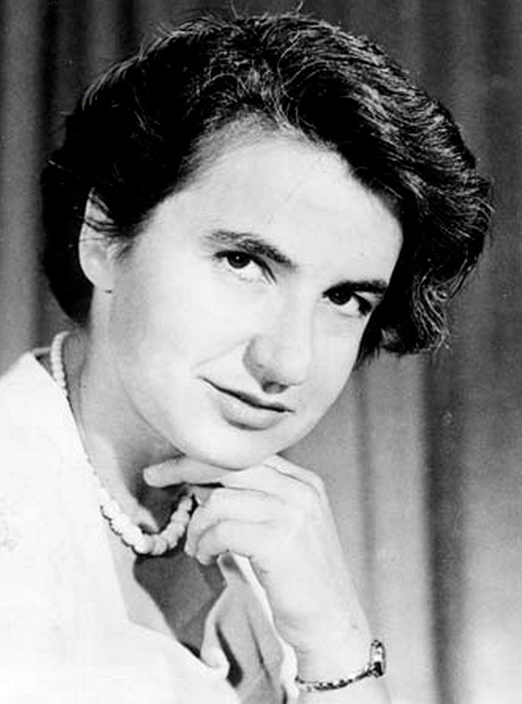
Rosalind Franklin’s draft reports on her theoretical models for DNA’s structure were key to Watson and Crick’s discovery of the double helix, but she received little recognition in her lifetime.
Making the pieces fit
By splitting the base quantities into two sets of pairs, Chargaff had shed light on the structure of DNA. Watson and Crick now began to think of adenine as only and always linking to thymine, and guanine to cytosine. In assembling the cardboard pieces for their 3-D jigsaw, Watson and Crick were juggling a vast amount of data, working from mathematics, X-ray images, their own knowledge of chemical bonds and their angles, and other data – all approximate and subject to ranges of errors. Their final breakthrough came when they realized that making slight adjustments to the configurations of thymine and guanine allowed the pieces to fit together, producing an elegant double helix in which the pairs of bases linked along the middle. Unlike the protein alpha helix, which had 3.6 sub-units in one complete turn, DNA had about 10.4 sub-units per turn.
The model that Watson and Crick described consisted of two helical or corkscrew phosphate-sugar backbones curling around each other, like the uprights of a “twisted ladder”, joined by pairs of bases serving as rungs. The sequence of bases worked like letters in a sentence, carrying small units of information that combined to make an overall instruction, or gene – which in turn told the cell how to make the particular protein or other molecule that was the physical manifestation of the genetic data and had a particular role in the cell’s fabric and function.
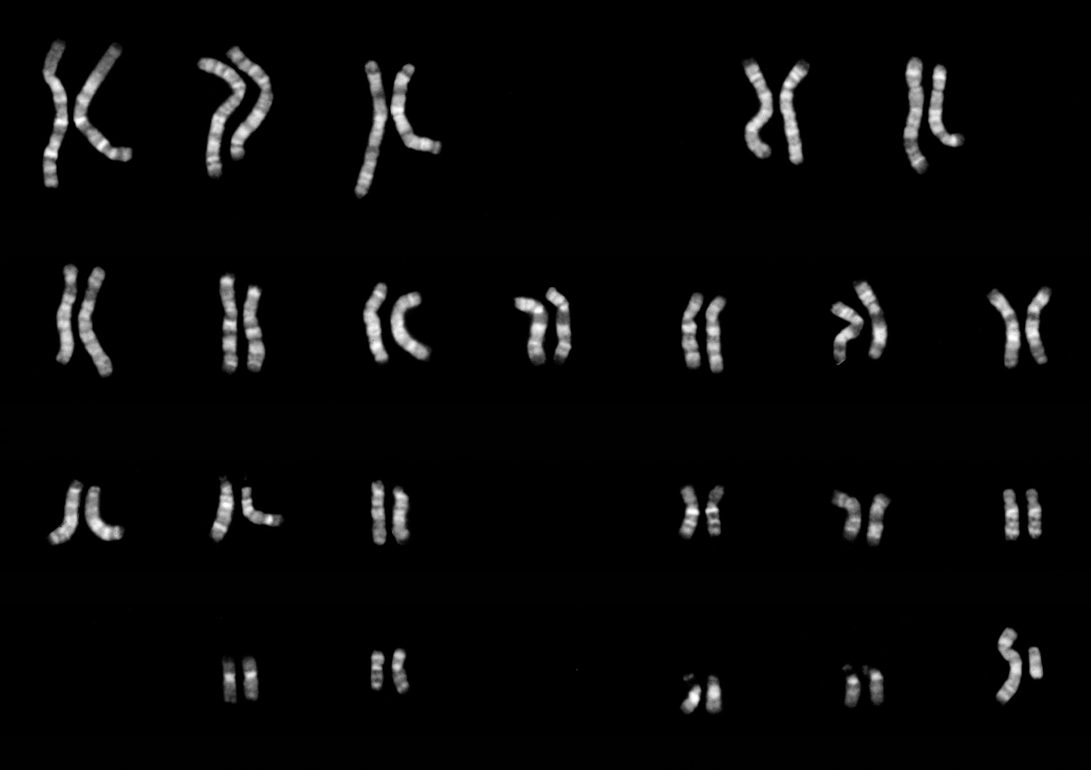
These are human male chromosomes. Before Crick and Watson’s discovery, it had been known that chromosomes carry genes that pass from a dividing cell to a daughter cell.
Zip and unzip
Each pair of bases is joined by what chemists call hydrogen bonds. These are made and broken relatively easily, so the sections of the double-helix can be “unzipped” by undoing the bonds, which then exposes the code of bases as a template for making a copy.
This zip-unzip allowed two processes to occur. First, a mirror complementary copy of nucleic acid could be made from one unzipped half of the double helix; then, carrying its genetic information as the sequence of bases, it would leave the cell nucleus to become involved in the protein production.
Second, when the whole length of the double helix was unzipped, each part would act as a template to build a new complementary partner – resulting in two lengths of DNA that were identical to the original and to each other. In this way, DNA was copied as cells divided into two for growth and repair throughout an organism’s life – and as sperm and eggs, the sex cells, carried their quotient of the genes to make a fertilized egg, so beginning the next generation.
“Secret of life”
On 28 February 1953, elated by their discovery, Watson and Crick went for lunch to The Eagle, one of Cambridge’s oldest inns, where colleagues from the Cavendish and other laboratories often met. Crick is said to have startled drinkers by announcing that he and Watson had discovered “the secret of life” – or so Watson later recalled in his book, The Double Helix, though Crick denied this really happened.
In 1962, Watson, Crick, and Wilkins were awarded the Nobel Prize in Physiology or Medicine “for their discoveries concerning the molecular structure of nucleic acids and its significance for information transfer in living material”. The award, however, was surrounded in controversy. In the preceding years, Rosalind Franklin had received little official credit for producing the key X-ray images and for writing the reports that helped to direct Watson and Crick’s research. She died of ovarian cancer in 1958, aged only 37, and was therefore ineligible for the Nobel Prize in 1962, since the prizes are not awarded posthumously. Some said the award should have been made earlier, with Franklin as one of the co-recipients, but the rules allow a maximum of three.
Following their momentous work, Watson and Crick became world celebrities. They continued their research in molecular biology and received great numbers of awards and honours. Now that the structure of DNA was known, the next big challenge was to solve the genetic “code”. By 1964, scientists worked out how sequences of its bases were translated into the amino acids that make up specific proteins and other molecules that are the building blocks of life.
Today, scientists can identify base sequences for all the genes of an organism, collectively known as its genome. They can manipulate DNA to move genes around, delete them from specific lengths of DNA, and insert them into others. In 2003, the Human Genome Project, the largest international biological research project ever, announced that it had completed the mapping of the human genome – a sequence of more than 20,000 genes. Crick and Watson’s discovery had paved the way for genetic engineering and gene therapy.
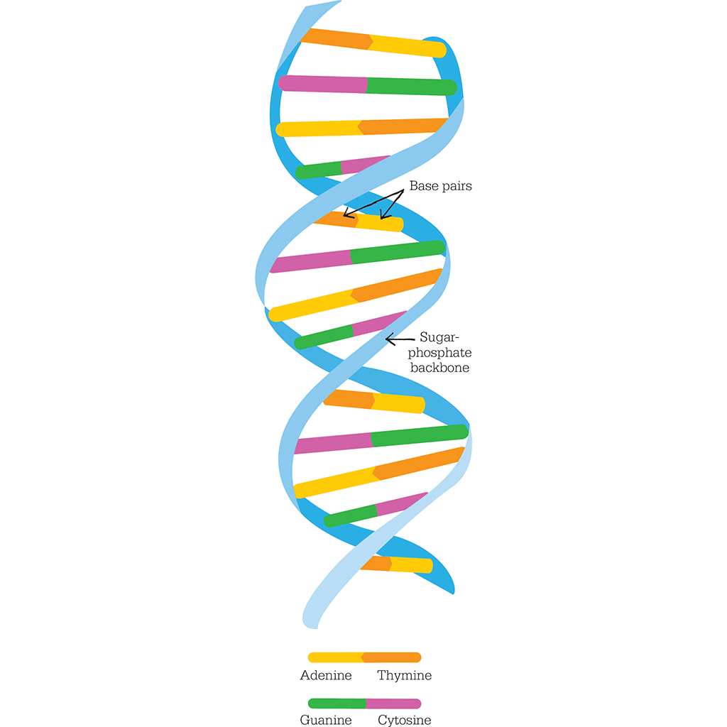
A DNA molecule is a double helix formed by base pairs attached to a backbone made of sugar-phosphates. The base pairs always match up in combinations of either adenine–thymine or cytosine–guanine.
"I never dreamed that in my lifetime my own genome would be sequenced."
James Watson
JAMES WATSON AND FRANCIS CRICK
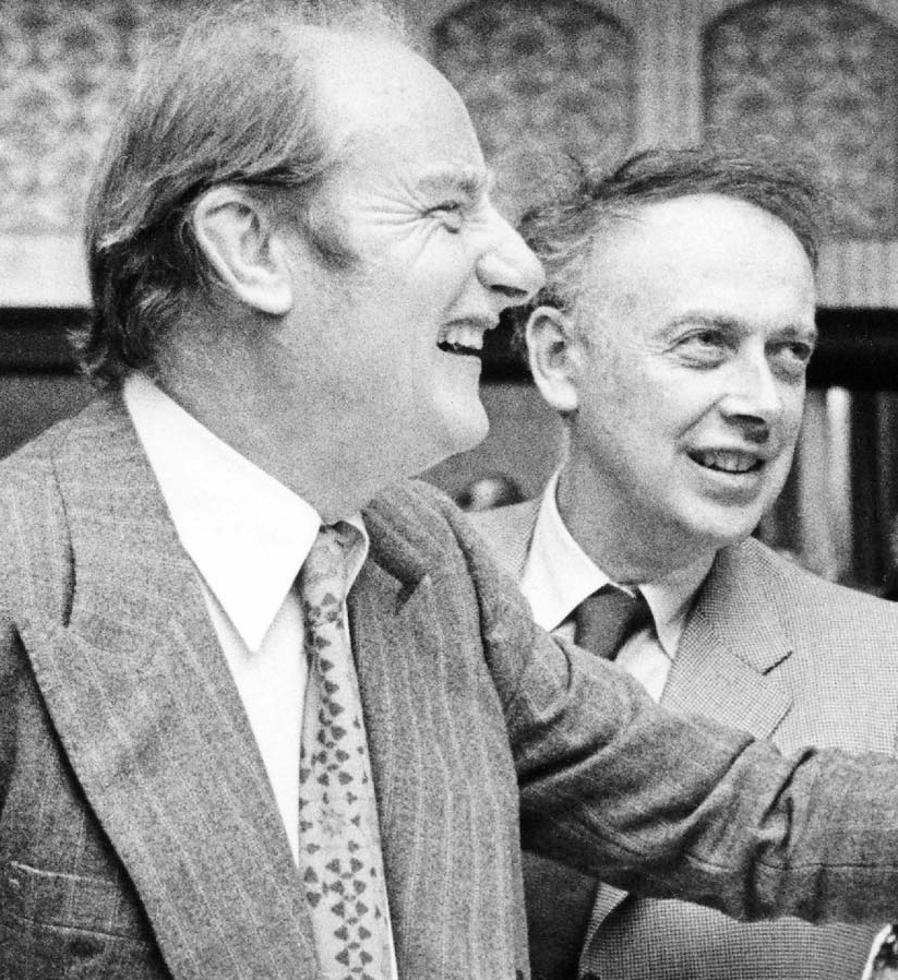
James Watson (on the right) was born in 1928 in Chicago, USA. At the precocious age of 15 he entered the University of Chicago. After postgraduate study in genetics, Watson moved to Cambridge, England, to team up with Francis Crick. He later returned to the USA to work at the Cold Spring Harbor Laboratory in New York. From 1988, he worked on the Human Genome Project, but left after a disagreement over patenting genetic data.
Francis Crick was born in 1916 near Northampton in Britain. He developed anti-submarine mines during World War II. In 1947, he went to Cambridge to study biology and here began work with James Watson. Later, Crick became known for the “central dogma”: that genetic data flow in cells in essentially one way. In later life, Crick turned to brain research and developed a theory of consciousness.
Key works
1953 Molecular Structure of Nucleic Acids: A Structure for Deoxyribose Nucleic Acid
1968 The Double Helix
See also: Charles Darwin • Gregor Mendel • Thomas Hunt Morgan • Barbara McClintock • Linus Pauling • Craig Venter