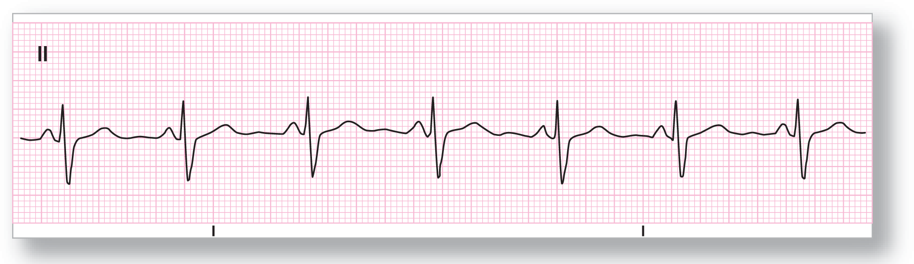
Figure 14-3 This rhythm strip is representative of the patient’s P-wave morphology and PR intervals while in normal sinus rhythm.
From Arrhythmia Recognition: The Art of Interpretation, courtesy of Tomas B. Garcia, MD.
Hint #1: Always Try to Compare Your New Strip to an Old ECG or Rhythm Strip
As you can imagine, the P-wave morphology in an ectopic atrial rhythm will differ from that found in normal sinus rhythm. (We discussed the origins of these morphologic differences in the ectopic P waves in Chapter 13, Premature Atrial Contraction. Quickly review these concepts again if you need to.) Many times the P waves are obviously ectopic in nature and easily identified. In some cases, however, the P-wave morphology can be very close to that of the normal sinus P-wave. In these cases, an old ECG or a strip showing the transition from normal sinus rhythm to the ectopic atrial pacemaker may be the only way to correctly identify this rhythm.
Take a look at Figure 14-2. This rhythm strip could easily be mistakenly diagnosed as normal sinus rhythm if viewed alone. Luckily, an old strip taken earlier of the same patient (Figure 14-3) was available. Notice the subtle, but significant, differences in the morphology of the P waves and the PR intervals between the two strips (Figure 14-4). Without the ability to compare the two strips, making the diagnosis would have been next to impossible.
Figure 14-3 This rhythm strip is representative of the patient’s P-wave morphology and PR intervals while in normal sinus rhythm.
From Arrhythmia Recognition: The Art of Interpretation, courtesy of Tomas B. Garcia, MD.
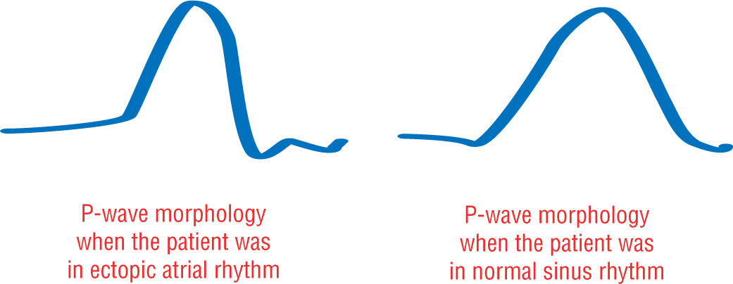
Figure 14-4 When the P-wave morphologies of Figure 14-2 and Figure 14-3 are magnified, the differences become obvious.
© Jones & Bartlett Learning.
Description