The Limbic System
We’ve considered the brain’s oldest parts, but we’ve not yet reached its newest and highest regions, the cerebral hemispheres (the two halves of the brain). Between the oldest and newest brain areas lies the limbic system (limbus means “border”). This system contains the amygdala, the hypothalamus, and the hippocampus (Figure 11.7).
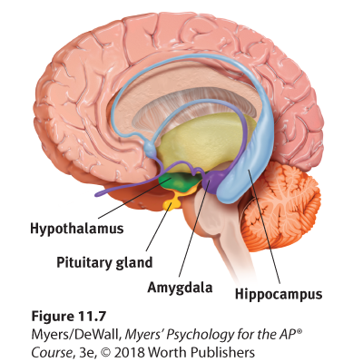
Figure 11.7 The limbic system
This neural system sits between the brain’s older parts and its cerebral hemispheres. The limbic system’s hypothalamus controls the nearby pituitary gland.
 Flip It Video: Limbic System
The Amygdala
Research has linked the amygdala, two lima-bean-sized neural clusters, to aggression and fear. In 1939, psychologist Heinrich Klüver and neurosurgeon Paul Bucy surgically removed a rhesus monkey’s amygdala, turning the normally ill-tempered animal into the most mellow of creatures. In studies with other wild animals, including the lynx, wolverine, and wild rat, researchers noted the same effect. So, too, with human patients. Those with amygdala lesions often display reduced arousal to fear- and anger-arousing stimuli (Berntson et al., 2011). One such woman, patient S. M., has been called “the woman with no fear,” even if being threatened with a gun (Feinstein et al., 2013).
What then might happen if we electrically stimulated the amygdala of a normally placid domestic animal, such as a cat? Do so in one spot and the cat prepares to attack, hissing with its back arched, its pupils dilated, its hair on end. Move the electrode only slightly within the amygdala, cage the cat with a small mouse, and now it cowers in terror.
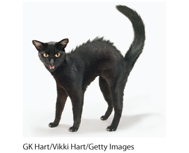
Aggression as a brain state Back arched and fur fluffed, this fierce cat is ready to attack. Electrical stimulation of a cat’s amygdala provokes angry reactions, suggesting the amygdala’s role in aggression. Which ANS division is activated by such stimulation?1
These and other experiments have confirmed the amygdala’s role in emotions such as fear and rage. One study found math anxiety associated with hyperactivity in the right amygdala (Young et al., 2012). Monkeys and humans with amygdala damage become less fearful of strangers (Harrison et al., 2015). Other studies link criminal behavior with amygdala dysfunction (Boccardi et al., 2011; Ermer et al., 2012). When people view angry and happy faces, only the angry ones increase activity in the amygdala (Mende-Siedlecki et al., 2013). But we must be careful. The brain is not neatly organized into structures that correspond to our behavior categories. When we feel afraid or act aggressively, there is neural activity in many areas of our brain—not just the amygdala. If you destroy a car’s battery, it’s true that you won’t be able to start the engine. But the battery is merely one link in an integrated system.
The Hypothalamus
Just below (hypo) the thalamus is the hypothalamus (Figure 11.8), an important link in the command chain governing bodily maintenance. Some neural clusters in the hypothalamus influence hunger; others regulate thirst, body temperature, and sexual behavior. Together, they help maintain a steady (homeostatic) internal state.
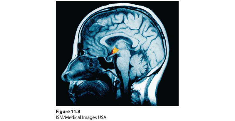
Figure 11.8 The hypothalamus
This small but important structure, colored yellow/orange in this MRI scan, helps keep the body’s internal environment in a steady state.
To monitor your body state, the hypothalamus tunes into your blood chemistry and any incoming orders from other brain parts. For example, picking up signals from your brain’s cerebral cortex that you are thinking about sex, your hypothalamus will secrete hormones. These hormones will in turn trigger the adjacent “master gland” of the endocrine system, your pituitary (see Figure 11.7), to influence your sex glands to release their hormones. These hormones will intensify the thoughts of sex in your cerebral cortex. (Once again, we see the interplay between the nervous and endocrine systems: The brain influences the endocrine system, which in turn influences the brain.)
A remarkable discovery about the hypothalamus illustrates how progress in science often occurs—when curious, open-minded investigators make an unexpected observation. Two young McGill University neuropsychologists, James Olds and Peter Milner (1954), were trying to implant an electrode in a rat’s reticular formation when they made a magnificent mistake: They placed the electrode incorrectly (Olds, 1975). Curiously, as if seeking more stimulation, the rat kept returning to the location where it had been stimulated by this misplaced electrode. On discovering that they had actually placed the device in a region of the hypothalamus, Olds and Milner realized they had stumbled upon a brain center that provides pleasurable rewards (Olds, 1975).
Later experiments located other “pleasure centers” (Olds, 1958). (What the rats actually experience only they know, and they aren’t telling. Rather than attribute human feelings to rats, today’s scientists refer to reward centers, not “pleasure centers.”) Just how rewarding are these reward centers? Enough to cause rats to self-stimulate these brain regions more than 1000 times per hour. Moreover, they would even cross an electrified floor that a starving rat would not cross to reach food (Figure 11.9).
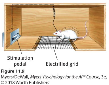
Figure 11.9 Rat with an implanted electrode
With an electrode implanted in a reward center of its hypothalamus, the rat readily crosses an electrified grid, accepting the painful shocks, to press a pedal that sends electrical impulses to that center.
In other species, including dolphins and monkeys, researchers later discovered other limbic system reward centers, such as the nucleus accumbens in front of the hypothalamus (Hamid et al., 2016). Animal research has also revealed both a general dopamine-related reward system and specific centers associated with the pleasures of eating, drinking, and sex. Animals, it seems, come equipped with built-in systems that reward activities essential to survival.
Researchers are experimenting with new ways of using brain stimulation to control nonhuman animals’ actions in search-and-rescue operations. By rewarding rats for turning left or right, one research team trained previously caged rats to navigate natural environments (Talwar et al., 2002; Figure 11.10). By pressing buttons on a laptop, the researchers were then able to direct the rat—which carried a receiver, power source, and video camera all in a tiny backpack—to turn on cue, climb trees, scurry along branches, and return.
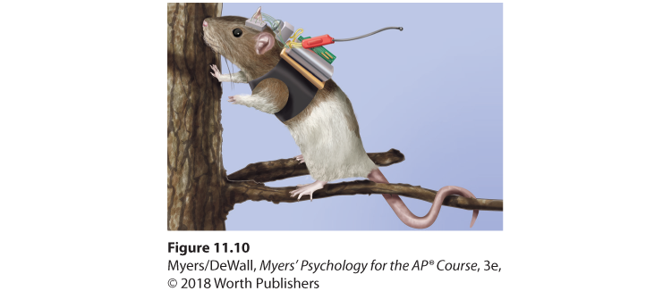
Figure 11.10 Ratbot on a pleasure cruise
When stimulated by remote control, a rat could be guided to navigate across a field and even up a tree.
Do humans have limbic centers for pleasure? To calm violent patients, one neurosurgeon implanted electrodes in such areas. Stimulated patients reported mild pleasure; unlike Olds’ rats, however, they were not driven to a frenzy (Deutsch, 1972; Hooper & Teresi, 1986). Moreover, newer research reveals that stimulating the brain’s “hedonic hotspots” (its reward circuits) produces more desire than pure enjoyment (Kringelbach & Berridge, 2012). Experiments have also revealed the effects of a dopamine-related reward system in people. For example, dopamine release produces our pleasurable “chills” response to a favorite piece of music (Zatorre & Salimpoor, 2013).
“ If you were designing a robot vehicle to walk into the future and survive, . . . you’d wire it up so that behavior that ensured the survival of the self or the species—like sex and eating—would be naturally reinforcing.”
Some researchers believe that substance use disorders may stem from malfunctions in natural brain systems for pleasure and well-being. People genetically predisposed to this reward deficiency syndrome may crave whatever provides that missing pleasure or relieves negative feelings (Blum et al., 1996).
The Hippocampus
The hippocampus—a seahorse-shaped brain structure—processes conscious, explicit memories and decreases in size and function as we grow older. Humans who lose their hippocampus to surgery or injury also lose their ability to form new memories of facts and events (Clark & Maguire, 2016). Those who survive a hippocampal brain tumor in childhood struggle to remember new information in adulthood (Jayakar et al., 2015). National Football League players who experience one or more loss-of-consciousness concussions may later have a shrunken hippocampus and poor memory (Strain et al., 2015). Module 54 offers additional detail about how hippocampus size and function decrease as we grow older. Module 32 explains how our two-track mind uses the hippocampus to process our memories.
* * *
Figure 11.11 locates the brain areas we’ve discussed, as well as the cerebral cortex, our next topic.
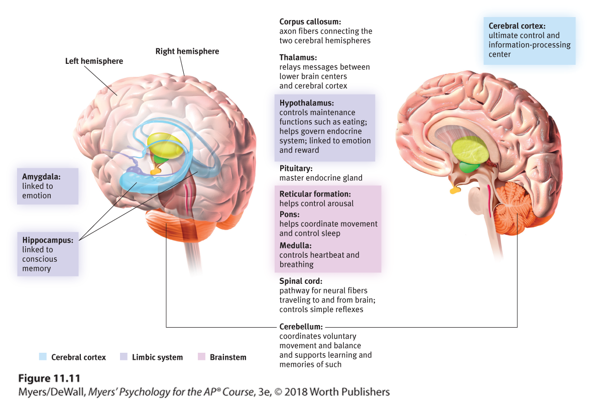
Figure 11.11 Brain structures and their functions