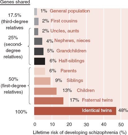
One of the most remarkable paradigm shifts that has occurred during our lives has been the recognition that most of our behavior is inherited: eccentric, compassionate, outgoing, irritable, and so on. Traits such as these travel in families from generation to generation. The same is true with many forms of mental illness. With schizophrenia, if one family member has the illness, the likelihood that a relative will also develop the disease increases as the percentage of shared DNA increases (Figure 1.1).
The domestication of animals provides another example of the genetic control of behavior. Charles Darwin, without any knowledge of genes, believed the temperament of domestic animals was inherited. Dmitry Belyaev, a Russian geneticist, validated this with his famous farm-fox experiment in Siberia.
Belyaev domesticated wild foxes simply by selecting and breeding the tamest animals. He started with 130 wild foxes and administered a little test of tameness. Animals were approached by humans. Those that were most tolerant were mated to each other and the process was repeated with the offspring. Within 20 years, the foxes were domesticated. Within 40 years, they were literally house pets. Furthermore, the domesticated foxes produced less corticosteroids (the stress hormones) and higher levels of serotonin than did control foxes.
But what role does the environment play in the development of our personalities? Bouchard and others have eloquently addressed this question by looking at personality characteristics in monozygotic (identical) and dizygotic (fraternal) twins—reared together and reared apart. Using personality tests to assess five major personality traits, they found more correlations for monozygotic twins compared with dizygotic twins regardless of whether they were raised together or apart (Table 1.1). In other words, the monozygotic twins reared apart shared more personality characteristics than did the dizygotic twins reared in the same household. Bouchard’s conclusion was that these personality traits are strongly influenced by inheritance and only modesty affected by environment. (The lay press unfortunately summarized this research as “Parents Don’t Matter.”)
The mean correlations are surprisingly similar to the lifetime risk of developing schizophrenia for identical (monozygotic) and fraternal (dizygotic) twins as shown in Figure 1.1.
Figure 1.2 shows a remarkable example of identical twins separated when 5 days old and raised in different households—one in Brooklyn and the other in New Jersey. They did not meet again until they were 31 years old. Both are firemen, bachelors, with mustaches, and metal frame glasses. Not only do they have the same mannerisms, but they laugh at the same jokes and enjoy the same hobbies. Yet, they were exposed to entirely different environmental influences throughout their lives. Our individuality—who we are, how we socialize, what we like, even our religious beliefs—is influenced more by the brain we are born with than by the experiences we have along the way.
An alternative perspective on the twin studies, however, makes us cautious about ignoring the role of environment on behavior. For example, even though there is about a 50% concurrence between monozygotic twins for schizophrenia (Figure 1.1) or extraversion (Table 1.1), the other 50% do not have the same condition—yet they share the same DNA. This is also true for bipolar disorder, alcoholism, and panic disorder. About half the monozygotic twins have the same illnesses while the other half do not.
FIGURE 1.1  As the shared genetic profile with someone having schizophrenia increases, the risk of developing schizophrenia also increases. (Adapted from Gottesman II. Schizophrenia Genesis. New York, NY: WH Freeman; 1991.)
Clearly, our brains are more programmed by our genes than we previously believed, but that does not explain everything. Experiences during our lives can affect the outcome, particularly trauma, and especially when the trauma occurs early in life. The challenge for us in trying to understand the brain and complex behaviors is to conceptualize and begin to unravel the brain mechanisms that are predetermined by genetics but can change in response to the environment.
BRIEF HISTORY OF NEUROSCIENCE
The brain has not always been of interest to humankind. Most ancient cultures did not consider the brain to be an important organ. Both the Bible and Talmud fail to mention diseases related to the central nervous system (CNS). Egyptians carefully embalmed the liver and the heart but had no use for the brain; they actually scooped it out and threw it away. (If there really is an Egyptian afterlife—those poor pharaohs are spending eternity without a brain.) So how did humans go from ignoring the brain to seeing it as the most complex organ in the universe?
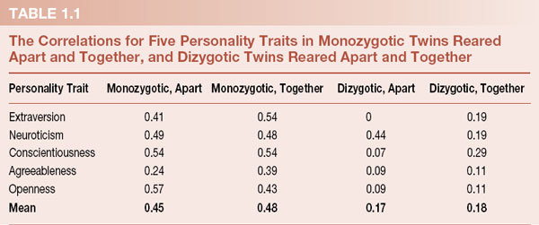
FIGURE 1.2  Gerald Levy (left) and Mark Newman are identical twins who were separated at birth, yet have made many of the same choices in life. (From The Image Works, Woodstock, New York.)
Thomas Willis—after whom the Circle of Willis at the base of the brain was named—was the first neurologist. In the 17th century, he moved us into what one author has called the Neurocentric Age. Before Willis—and actually for a considerable time after him—physicians based their understandings of illness on the writings of the great physicians from antiquity. Willis took the unusual approach of describing a patient’s behavior, then examining the brain after death and making correlations.
He was the first to coin terms such as lobe, hemisphere, and corpus striatum—terms that we still use. Comparing the human CNS anatomy with that of animals and conducting postmortem dissection of interesting cases, he made surprisingly accurate conclusions about higher brain functions versus lower brain functions. For example, he deduced that human functions such as memory were likely to reside in the “outmost banks” (gray matter) of the cerebral hemispheres because these areas were smaller in animals and damaged in individuals with severe head injuries who had lost memory. He believed that the brainstem likely controlled basic functions such as breathing and heart rate. However, he also thought that the white matter was the seat of imagination. He was not entirely on target, but he started the process of matching structures with behaviors.
At the start of the 18th century, it was still not clear how nerves transmitted information. Luigi Galvani, an Italian physician (memorialized by the term galvanic skin response), demonstrated through extensive experimentation that a frog muscle would twitch when stimulated with electricity. This established that the substance flowing through the nerves was not air, fluid, or spirits, but electricity. Galvani proposed that the brain secretes electricity, which is then distributed to the muscles by the nerves. He also believed that the electricity did not leak into the surrounding tissue because the nerves were covered with a fatty insulation, which we now know is myelin. Not all his beliefs were accurate, but he made the big leap to recognizing that muscle movement in mammals is stimulated by intrinsic electrical activity.
Before the 1860s the brain was seen as a single multipurpose organ, much the way we currently view the liver or pancreas. The French physician Paul Broca with his famous case in 1861 confirmed for the first time that certain functions were localized to specific regions of the brain (Figure 1.3). The patient exhibited a loss of articulate speech. All he could express was one syllable: “tan.” His utterances could convey great emotional tone, but the syllable never changed. However, he retained oral dexterity, and he could hear and comprehend. After his death, an autopsy revealed a lesion of his left frontal lobe—what is now called Broca’s aphasia. The fact that almost all similar cases were on the left hemisphere and that similar right hemispheric lesions did not affect speech also led Broca to identify left/right dominance for some functions.
David Ferrier of Scotland and Eduard Hitzig of Germany independently identified the specialized cortical areas controlling motor function. Using the techniques of stimulation and ablation in experimental animals, they localized and mapped out what we now call the motor cortex. This new understanding of the brain provided the first examples of useful neurosurgical treatment given on the basis of the patient’s motor symptoms. There is a case reported from 1879 of a teenage girl with seizures of the right face and arm who had a left meningioma accurately diagnosed and removed. With the development of antiseptic techniques and effective anesthesia, surgeons could successfully localize and remove some tumors using Ferrier’s map of the motor cortex.
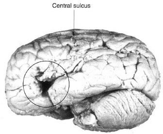
FIGURE 1.3  The preserved brain of the patient who helped Broca convince physicians that some functions—in this case the ability to speak—were localized in the cerebrum. (Adapted from Bear MF, Connors BW, Paradiso MA, eds. Neuroscience: Exploring the Brain. 3rd ed. Baltimore, MD: Lippincott Williams & Wilkins; 2007.)
It is noteworthy that both men speculated about higher brain functions and the lobes in front of the motor cortex—the prefrontal cortex. Ferrier noted problems with attention in monkeys with damaged frontal lobes. Experimenting with dogs, Hitzig came to believe that the frontal cortex played an important role in abstract thought.
The discovery of individual neurons was a major step in the development of neuroscience. In order to be able to see nerve cells, it was necessary to be able to fix the brain (which can have the consistency of gelatin) and to cut thin slices; in addition, microscopes of sufficient power were needed. An Italian physician, Camillo Golgi, discovered a selective silver stain that allowed researchers to visualize the individual nerve cells in what was otherwise a uniform blob of color. For the first time, researchers could see sharp black images of the nerve cells and identify specific parts such as the cell body and the dendritic branches (Figure 1.4). Termed the Golgi stain, this technique is still used today.
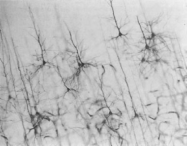
FIGURE 1.4  Pyramidal nerve cells after incubation with Golgi’s silver stain. Only about 1% of the neurons absorb the stain, which allows for the identification of individual cells in what would otherwise be a very crowded slice. (Adapted from Bear MF, Connors BW, Paradiso MA, eds. Neuroscience: Exploring the Brain. 3rd ed. Baltimore, MD: Lippincott Williams & Wilkins; 2007.)
Santiago Ramón y Cajal, a Spanish physician, used Golgi’s stain, a Zeiss light microscope, and 25 years of patient observation to become perhaps the first modern neuroscientist. He proposed that individual nerve cells are the singular unit of the brain—a new concept at that time, which has since been called the neuron doctrine. By tracing neurons from sensory organs such as the eye back to the cortex and from the motor cortex to the muscles, he concluded that the dendrites are receptive, the cell body is executive, and the axon transmits the information over a long distance.
Ramón y Cajal went on to show that nerve impulses flow only in one direction—what he called his law of dynamic polarization. Figure 1.5 is one of his drawings, with little arrows showing the direction of the impulses between two communicating neurons. Additionally, Ramón y Cajal observed and meticulously drew the embryonic development of neurons. He was the first to document the growth of an axon that ultimately branches with dendrites and collateral axons. In 1894, Ramón y Cajal stated in a lecture to the Royal Society of London, “the ability of neurons to grow in an adult and their power to create new connections can explain learning.” This is often cited as the origin of the synaptic theory of memory. Both Golgi and Ramón y Cajal received the Nobel Prize for Physiology and Medicine in 1906.

FIGURE 1.5  Drawing of neurons by Ramón y Cajal showing the unidirectional nature of impulses in the communication between neurons A and B. (From Ramón y Cajal S. Recollections on My Life. Transactions of the American Philosophical Society. Vol. 8, Part 2. Philadelphia, PA: The American Philosophical Society; 1937.)
Charles Sherrington, an English neurophysiologist, did extensive studies of animal nerves. Focusing mostly on the spinal and peripheral nerves, he advanced the understanding of sensory dermatomes and reflexes. He is best known for giving us the term synapse—although he never actually saw one. Sherrington theorized that a physical junction existed between nerves to pass along an impulse. The synapse would not be seen until the development of the electron microscope in the 1950s.
Another English neurophysiologist (who shared the 1932 Nobel Prize with Sherrington), Edgar Adrian, is best known for recognizing the “all-or-nothing” properties of the action potentials. With regard to behavior and the brain, his major discoveries dealt with habituation and the sensory cortex. Adrian found that a stimulus to a nerve cell is followed by a burst of action potentials coursing down the axon. However, the quantity of action potentials decreases over time even if the stimulus remains unchanged. This may explain at the level of the neuron the basis for a well-known treatment for anxiety: exposure therapy.
In the beginning of the 20th century, it was not known how neurons communicate with each other or how a neuron can make a muscle contract. Some believed the communication was electrical—as though a spark jumped from one neuron to another. Others believed that a chemical process transmitted the signal. No one had evidence establishing one system or another. Henry Dale and Otto Loewi, an Englishman and a German, shared the Nobel Prize in 1936 for their work in establishing the chemical transmission of nerve impulses. Dale was working with the autonomic nervous system and found that an epinephrine-like compound had activating effects on the sympathetic nervous system, and that acetylcholine could activate the parasympathetic nervous system as well as skeletal muscles. Unfortunately, Dale was unable to show that epinephrine (or really norepinephrine) and acetylcholine were excreted by the neurons to elicit these effects.
It was Otto Loewi who in 1921 performed the elegant little experiment that proved the neurochemical transmission of nerve impulses. Legend has it that Loewi dreamed the experiment and, upon awaking early in the morning, rushed down to the laboratory and performed it. The experiment is depicted in Figure 1.6. Loewi’s clever experiment showed that stimulating the vagus nerve slowed down the beating of the frog heart bathed in Ringer’s solution. He then transferred some of that solution to another isolated frog heart and, without electrical stimulation, its rhythm also slowed down—as though its vagus nerve had been stimulated. He concluded that a chemical was excreted from the synapses of the first heart when the vagal nerve was stimulated. This chemical then flowed into the container holding the second heart and induced bradycardia.
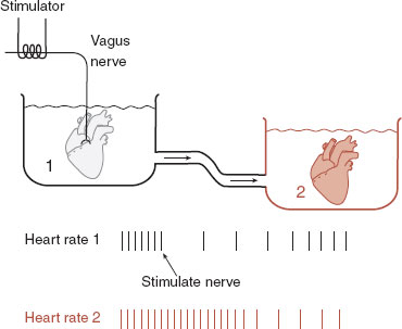
FIGURE 1.6  Otto Loewi’s famous experiment establishing that a chemical from the vagus nerve of one heart can induce bradycardia in a second unstimulated heart.
In 1939, Hodgkin and Huxley published the first intracellular recording of an action potential (Figure 1.7). Before this time, no one had directly measured the electrical charge in an axon as an action potential passed. Hodgkin and Huxley were able to accomplish this by inserting microelectrodes into the giant axons of squids.
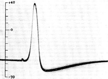
FIGURE 1.7  First published intracellular recording of an action potential. (From Hodgkin AL, Huxley AF. Action potentials recorded from inside a nerve fibre. Nature. 1939;144:710-711.)
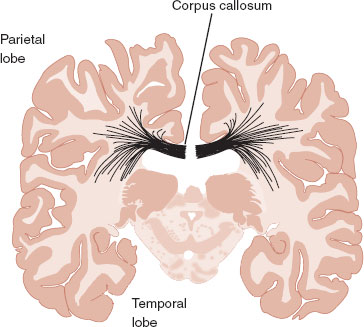
FIGURE 1.8  The corpus callosum contains millions of axons crossing between the two hemispheres. Sperry established that surgical resection of this structure interrupts the passage of information from one hemisphere to another.
By the 1940s, neurosurgeons were transecting the corpus callosum for a few patients with intractable seizures (Figure 1.8). The procedure prevented the spread of electrical activity across the corpus callosum into the opposite hemisphere and often provided significant relief for the patient. Remarkably, the patient’s personality and intellectual functioning appeared unaffected by the drastic procedure. Robert Sperry, with careful experimentation, established that the left and right hemispheres no longer share information after this procedure. Furthermore, he was able to prove that the hemispheres have different functions. The left hemisphere is more involved with linear reasoning, language, and routine. The right hemisphere controls language intonation, spatial orientation, and processing novel situations.
Sperry won the 1981 Noble Prize in Medicine for his work with “split-brain” research. He shared the prize with David Hubel and Torsten Wiesel (see Chapter 6 for more on Hubel and Wiesel).
The CNS contains small quantities of proteins that some people call “brain fertilizer.” It is an apt description, as these molecules facilitate the growth of nerve cells. Rita Levi-Montalcini and her colleagues accidentally discovered the first of this class of proteins, now called nerve growth factors (NGFs) or trophic factors. They observed sensory nerve cells exploding with growth when cultured next to a mouse sarcoma. Recognizing the significance of their discovery, they painstakingly isolated the protein that they called as nerve growth factor. Figure 1.9 shows the profound impact NGF has on the growth of sensory neurons. Since this landmark discovery, many other growth factors have been isolated. Some of these growth factors, or the absence of these growth factors, appear to play important roles in how the brain develops and changes with experience. They are thus key targets in trying to understand many psychiatric diseases.
Without a doubt, proving that the adult brain can change in response to experience (plasticity) has been the most exciting discovery in neuroscience. While many individuals have been involved in this research, no single individual has done more than Eric Kandel. Kandel was able to prove what Cajal could only speculate about—that learning changes the cells of the brain, and even the chemical composition of those cells. Kandel worked with the simple sea snail Aplysia because it can remember and has only about 20,000 neurons in its entire CNS (compared with 100 billion in humans). Kandel taught the snail a few simple tasks—habituation (gradually ignoring an innocuous stimuli) and sensitization (remembering an aversive stimuli) (Figure 1.10A).
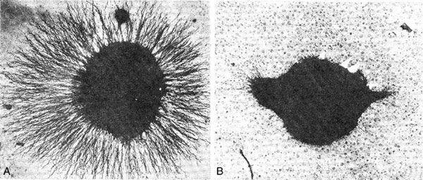
FIGURE 1.9  The sensory nerves in (A) show robust growth when exposed to nerve growth factor. (From Levi-Montalcini R. The nerve growth factor. Ann NY Acad Sci. 1964;118:149-170.)
For Aplysia, the gill with its siphon is a sensitive and important organ—one that it quickly withdraws with any sign of danger. Habituation is induced by sequentially touching the siphon with a soft brush. With repetition, the snail learns to ignore the gentle stimuli. Sensitization, on the other hand, is elicited with an electrical shock to the tail. This is something not to forget. Indeed, after many sessions the gill is still retracted with great vigor. After teaching Aplysia these skills, Kandel and his colleagues dissected and analyzed the changes in the sensory neurons. With habituation, the neurons regressed while with sensitization the number and size of the synaptic terminals grew (Figure 1.10B). This study and others like it are serving as the basic science building blocks for better understanding normal behavior and mental illness.
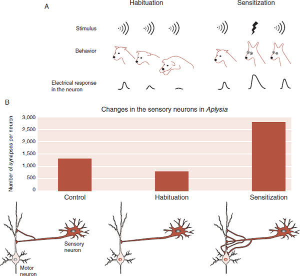
FIGURE 1.10  A. Animals learn to ignore a benign stimulus but continue to react to noxious stimuli. B. Cellular changes occurred in Aplysia with habituation and sensitization. (Adapted from Kandel ER. In Search of Memory. The Emergence of a New Science of Mind. New York, NY: W. W. Norton & Co.; 2006.)
Imaging
Researchers such as Willis and Broca were forced to wait for a patient to die before they could examine the brain. These scientists were studying patients with damaged brains using what some have called the lesion method. Although this remains an important tool for the modern neuroscientist (now there are large “brain banks” preserving brains of patients with similar illnesses), the noninvasive analysis of the CNS has transformed the way we study behavior and mental disorders.
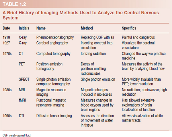
Early attempts to image the brain were unhelpful, painful, and even dangerous. An ordinary x-ray provides little information because the brain is soft tissue and not radiopaque. Searching for the displacement of calcified structures could provide indirect evidence of a mass. Pneumoencephalography, in which cerebrospinal fluid is removed and replaced with air to enhance visualization of the CNS, is an example of the painful and dangerous extremes that were foisted on patients in earlier times.
The development of noninvasive imaging techniques (Table 1.2) has led to another small revolution in neuroscience. Although the functional studies (positron emission tomography, single photon emission computerized tomography [SPECT], and functional magnetic resonance imaging) remain largely limited to research, the noninvasive structural analyses (computed tomography and magnetic resonance imaging [MRI]) have transformed the practice of neurology. Diffusion tensor imaging, a technique using MRI scans to measure the movement of water in tissue, creates images of white matter tracts.
A word of caution regarding brain imaging studies and psychiatric disorders. Throughout this book, we mention numerous studies examining brain volume or brain function for various psychiatric disorders. However, Ioannidis recently reviewed brain volume studies and found that the reported number of positive studies far exceeded what would be expected based on the power estimates of the studies. He believes the studies that do not find a significant difference between control subjects and patients are seldom published. The Ioannidis review reminds us that, in spite of the wonderful images of the brain shown in these studies, these studies may be misleading.
The figure shows a method of using imaging studies frequently found in the literature. That is, one functional study is subtracted from another and the result is superimposed on a structural image. In this case, the subject is performing a finger opposition task with his right fingers while in a SPECT scanner (A). The white arrow shows the activation of the left motor cortex. B. A SPECT scan in the controlled state (not moving) is also produced. C. The control image is subtracted from the task image. D. The results are superimposed on an MRI of the same location, and drawings of the human homunculus along the motor cortex are added for further understanding.

Functional imaging subtraction study superimposed on a structural image.
Nonhuman Animal Studies
Nonhuman animal studies provide another technique for understanding the marvels of the brain. The nonhuman animal brain is accessible in ways that are beyond the ethics of human research. Although they might have paws and whiskers, our human brains have much in common with those of nonhumans. The protein-coding regions of the mouse and human genomes are 85% identical. Nature is conservative and many of the molecular and cellular mechanisms that underlie the behavior are preserved from one species to the next. However, animals do not possess a similarly developed human cerebral cortex, nor can we ever be sure they actually have the psychiatric symptoms being studied. Besides the well-known microelectrode stimulation or ablation studies, there are several modern techniques that can be used on nonhuman brains that are worth reviewing.
Markers of Gene Activation
Two words of advice we would like to give to any student interested in neuroscience: gene expression. The DNA that gets turned on (or turned off) is called “gene expression” and this then controls the growth and activity of the brain. Understanding which genes are responsible is the key to understanding the brain and behavior. Researchers can now measure mRNA or proteins that result from gene expression. Cyclic adenosine monophosphate responsive element binding (CREB) protein and proteins in the Fos family are two transcription factors that are frequently used as markers of gene expression. Identifying CREB or FOS in a postmortem brain slice helps pinpoint the areas in the brain that were active in the animal during the experimental manipulation.
Knockout Mice
Animals (typically mice) can be engineered so that certain genes are turned off. Animals with silenced genes are called knockout mice. They are raised (if possible) and observed for changes in behavior compared with control mice—often called “wild” mice. Knockout mice have been used to understand obesity, substance abuse, and anxiety. While these studies represent a valuable research tool, one has to be cautious about generalizing from the results. The downstream effects of silencing the gene during development can never be fully appreciated.
Transgenic Mice
Transgenic mice are genetically engineered creatures. DNA from one organism is introduced into the DNA of a mouse egg, which is then fertilized. The adult mouse incorporates the foreign DNA into its genome. For example, DNA from jellyfish encoding for fluorescent proteins has been inserted into the mouse genome. Brain slices from these mice “light up” when viewed under fluorescent microscopes. Likewise, the ability to insert disease-causing DNA into mice and then observe the damage it causes to the brain has revolutionized neurology.

FIGURE 1.11  The microarray chip contains multiple copies of many different genes so that a broad spectrum of gene activity can be analyzed quickly in a scanner. (Adapted from Friend SH, Stoughton RB. The magic of microarrays. Sci Am. 2002;286:44-53.)
Viral-Mediated Gene Transfer
Viruses can be used as a vehicle to insert a section of DNA into the brain of living animals at specific locations. When the DNA is incorporated into the host DNA, new genes are expressed with possible alterations in behavior. For example, a virus was used to implant the DNA for the vasopressin receptor in the ventral pallidum of promiscuous voles. Responding voles were transformed into monogamous, family-oriented, church-going, and card-carrying conservatives (see Chapter 14 for details).
DNA Microarrays—Also Called Gene Chips
DNA microarrays enable researchers to compare the mRNA (and therefore gene activity) found in a tissue sample with the DNA of known identity. The microarray is a chip no bigger than a postage stamp with thousands of different DNA molecules, multiplied, segregated, and attached in separate tiny locations (Figure 1.11).
The mRNA from the tissue being studied is transcribed to DNA, labeled with fluorescent markers, and dropped onto the microarray chip. The single-stranded DNA from the tissue sample will bind with similar single-stranded DNA on the microarray. The chip is then read in a scanner that calculates the amount of binding between the tissue DNA and the chip DNA in each discrete spot, giving an estimate of that specific gene activity in the tissue. As an example, this procedure was done with small samples from the prefrontal cortex of schizophrenic and control postmortem brain. The schizophrenic brains showed reduced expression of myelination-related genes, suggesting a disruption in the myelin as part of the pathogenesis of schizophrenia (see Chapter 23).
The Controlled Trial
It is discouraging to realize that the brain is so resistant to change. It is more discouraging to read about eccentric clinicians, parents, teachers, and other meddlers expounding the effectiveness of their unproven interventions to reduce symptoms or improve behavior. Sugar and hyperactivity are “known” to have a cause and effect relationship that has unfortunately failed to materialize in controlled trials.
We tend to conceptualize the pathogenesis of psychiatric disorders as the absence of what we are replacing with our treatment (neurotransmitters, a superego, the corrective emotional experience, etc.). It is important to prove that these interventions are actually working. Perhaps, the greatest research tool in health care has been the controlled clinical trial. With this technique we can determine with some confidence how effective interventions might be, which then gives us some insight into the working of the brain.
Part 1: Match the names in the right column with the events in the left column
1. Started the Neurocentric Age |
A. Edgar Adrian |
2. The brain has intrinsic electric activity |
B. Paul Broca |
3. Localization of function |
C. Santiago Ramón y Cajal |
4. The motor cortex |
D. David Ferrier |
5. Silver stain |
E. Luigi Galvani |
6. Individual nerve cells are the singular unit of the CNS |
F. Camillo Golgi |
7. Coined the term synapse |
G. Hodgkin and Huxley |
8. “All-or-nothing” |
H. Otto Loewi |
9. Neurochemical transmission of nerve impulses |
I. Rita Levi-Montalcini |
10. First action potential |
J. Charles Sherrington |
11. Chemical guidance of regenerating nerves |
K. Robert Sperry |
12. Nerve growth factors |
L. Thomas Willis |
Part 2: Match the columns
13. Implanted micropipette |
M. DNA microarray |
14. Activation of DNA |
N. Gene expression |
15. Missing receptors |
O. Knockout mice |
16. Single-stranded DNA |
P. Microdialysis |
17. Implanting specific traits |
Q. Viral-mediated gene transfer |
See Answers section at the end of the book.