16 Neurovasculature
Arteries of the Abdomen
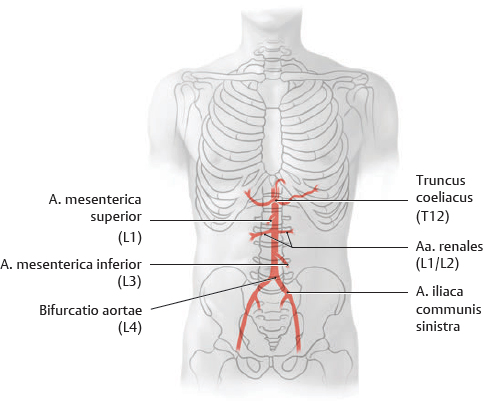
Fig. 16.1 Pars abdominalis aortae and major branches
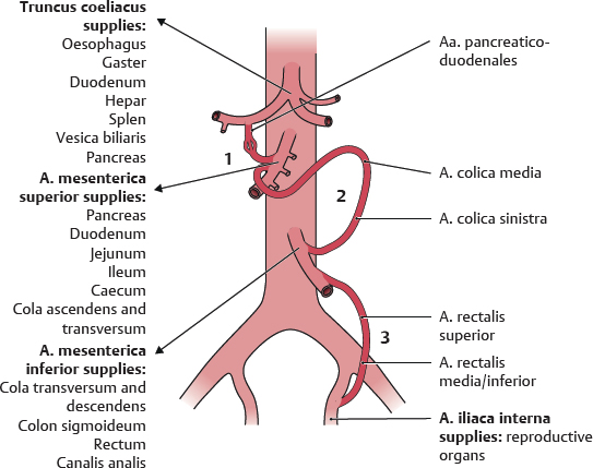
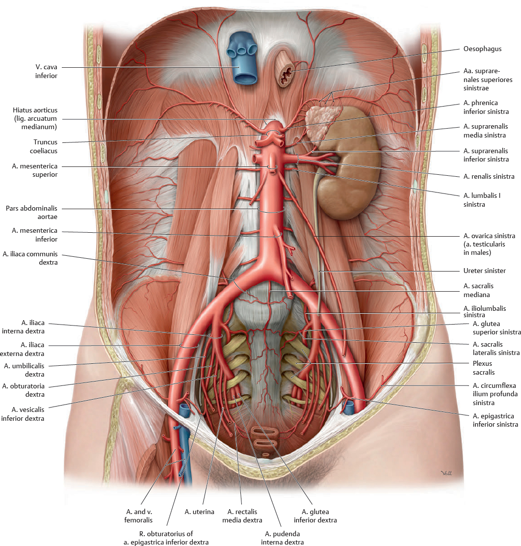
Anterior view. The pars abdominalis aortae enters the abdomen at the T12 level through the hiatus aorticus of the diaphragma (see p. 66). Before bifurcating at L4 into its terminal branches, the aa. iliacae communes, the pars abdominalis aortae gives off the aa. renales (see p. 183) and three major trunks that supply the organs of the gastrointestinal system:
Truncus coeliacus: Supplies the structures of the praeenteron (foregut), the anterior portion of the alimentary canal. The praeenteron consists of the oesophagus (abdominal 1.25 cm), gaster, duodenum (proximal half), hepar, vesica biliaris, and pancreas (superior portion).
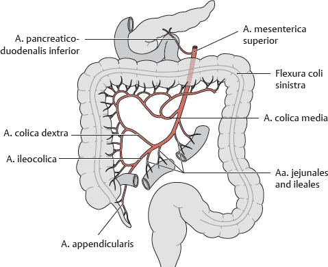
Arteria mesenterica superior: Supplies the structures of the mesenteron (midgut): the duodenum (distal half), jejunum and ileum, caecum and appendix vermiformis, colon ascendens, flexura coli dextra, and the proximal half of the colon transversum.
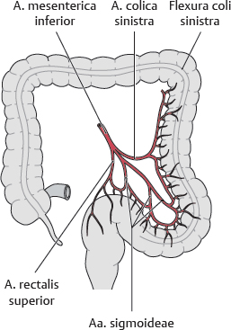
Arteria mesenterica inferior: Supplies the structures of the metenteron (hindgut): the colon transversum (distal half), flexura coli sinistra, cola descendens and sigmoideum, rectum, and canalis analis (upper part).
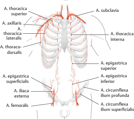
Fig. 16.2 Arteries of the abdominal wall
The aa. epigastricae superiores and inferiores form a potential anastomosis, or bypass for blood, from the aa. subclavia and femoralis. This effectively allows blood to potentially bypass the pars abdominalis aortae.
Fig. 16.3 Truncus coeliacus
Fig. 16.4 Arteria mesenterica superior
Fig. 16.5 Arteria mesenterica inferior
Fig. 16.6 Abdominal arterial anastomoses
Three major anastomoses provide overlap in the arterial supply to abdominal areas to ensure adequate blood flow. 1–Between the truncus coeliacus and the a. mesenterica superior via the aa. pancreaticoduodenales. 2–Between the a. mesenterica superior and a. mesenterica inferior via the a. colica media and a. colica sinistra. 3–Between the a. mesenterica inferior and the a. iliaca interna via the aa. rectales superior and media or inferior.
Pars Abdominalis Aortae & Arteriae Renales
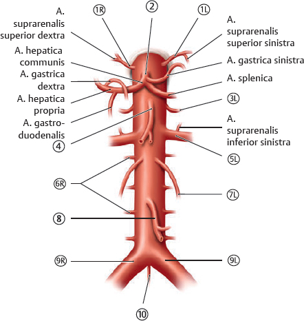
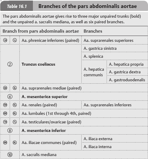
Fig. 16.7 Pars abdominalis aortae
Anterior view of the female abdomen. Removed: All organs except the left ren and gl. suprarenalis. The pars abdominalis aortae is the distal continuation of the pars thoracica aortae (see p. 80). It enters the abdomen at the T12 level and bifurcates into the aa. iliacae communes at L4.
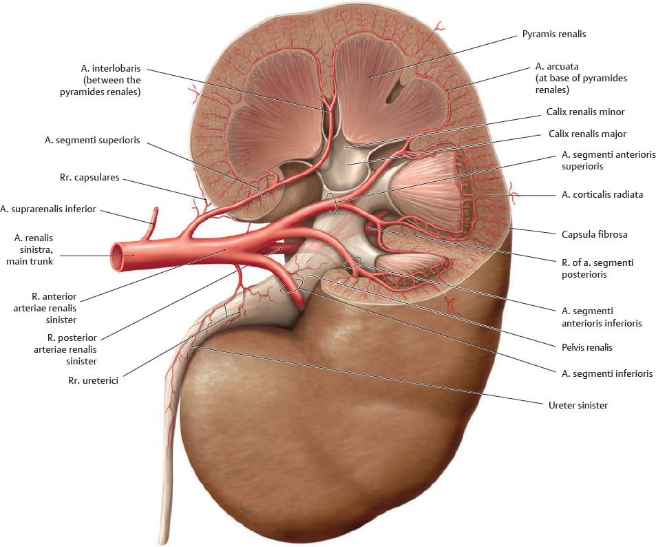
Fig. 16.8 Arteriae renales
Left ren, anterior view. The aa. renales arise at approximately the level of L2. Each a. renalis divides into a r. anterior and a r. posterior. The r. anterior further divides into four aa. segmentorum (circled).
 Clinical box 16.1
Renal hypertension
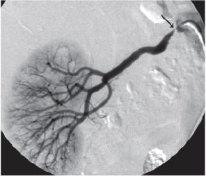
The ren is an important blood pressure sensor and regulator. Stenosis (narrowing) of the a. renalis reduces blood flow through the ren and stimulates increased production of renin, an enzyme that cleaves angiotensinogen to form angiotensin I. Subsequent cleavage yields angiotensin II, which induces vasoconstriction and an increase in blood pressure. Renal hypertension must be excluded (or confirmed) when diagnosing high blood pressure.
Stenosis of the right a. renalis (arrow), visible via arteriography.
Truncus Coeliacus
 The distribution of the truncus coeliacus is shown on p. 185.
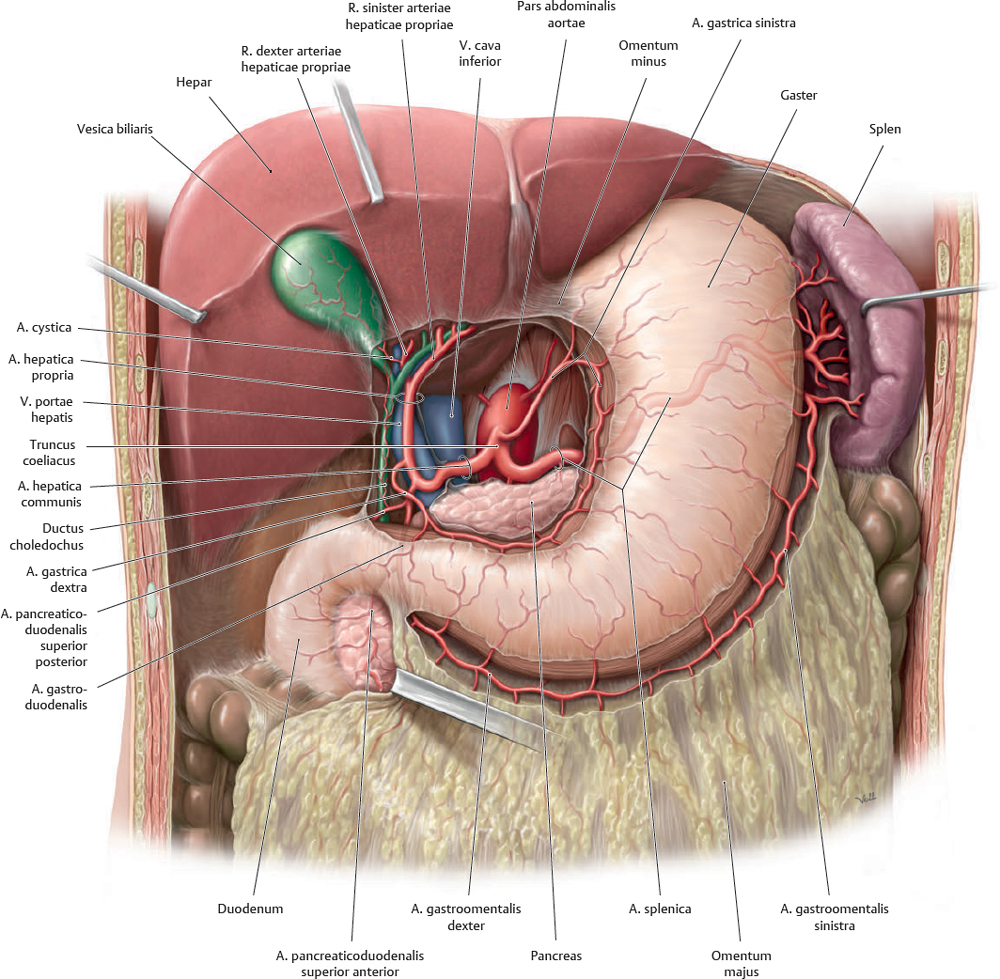
Fig. 16.9 Truncus coeliacus: Gaster, hepar, and vesica biliaris
Anterior view. Opened: Omentum minus. Incised: Omentum majus. The truncus coeliacus arises from the pars abdominalis aortae at about the level of T12/L1.
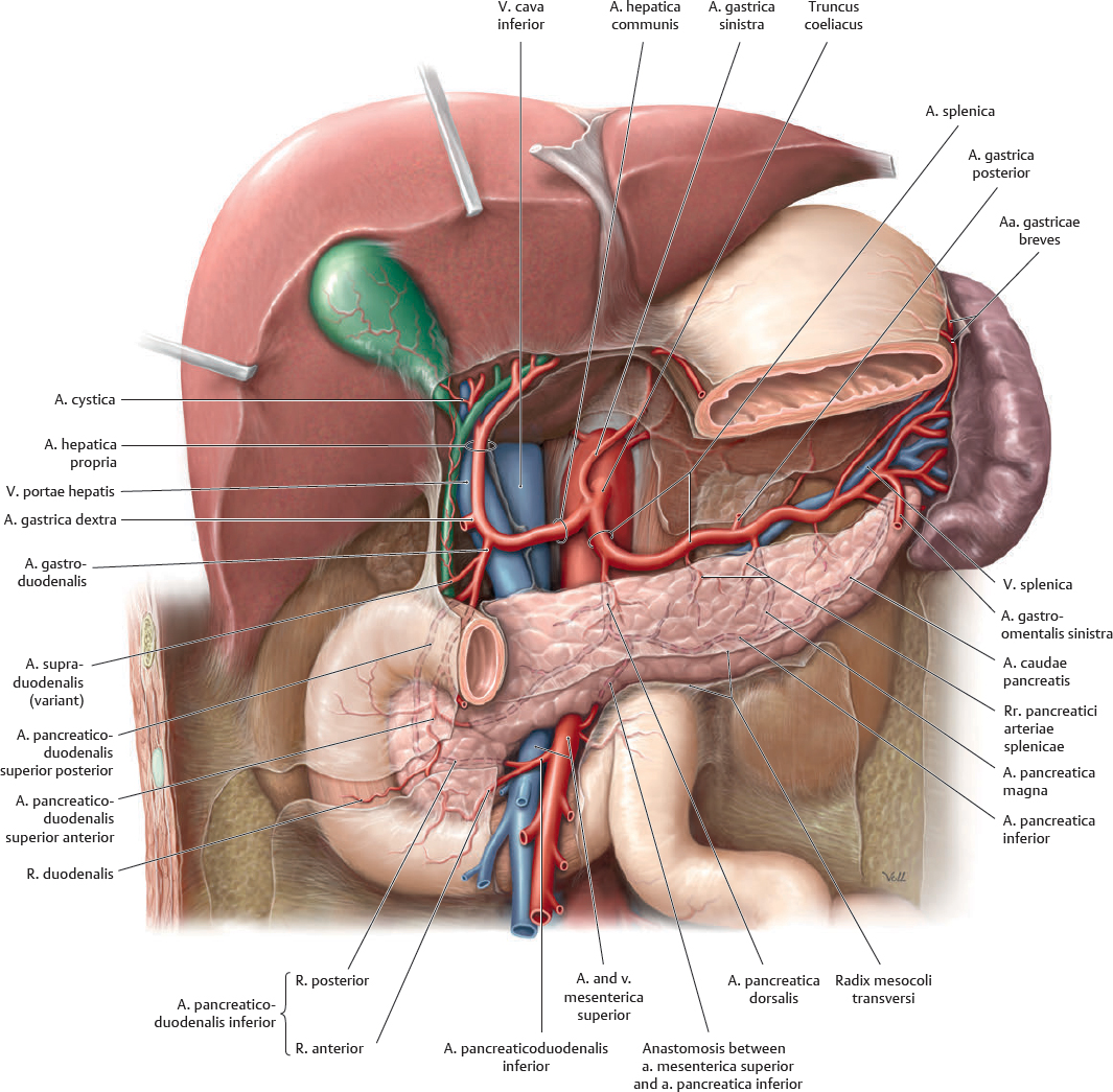
Fig. 16.10 Truncus coeliacus: Pancreas, duodenum, and splen
Anterior view. Removed: Corpus gastricum and omentum minus.
Arteria Mesenterica Superior & Arteria Mesenterica Inferior
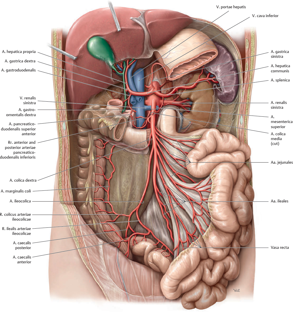
Fig. 16.11 Arteria mesenterica superior
Anterior view. Partially removed: Gaster, duodenum, and peritoneum. Reflected: Hepar and vesica biliaris. Note: The a. colica media has been truncated (see Fig. 16.12). The a. mesenterica superior and a. mesenterica inferior arise from the aorta opposite L1 and L3, respectively.
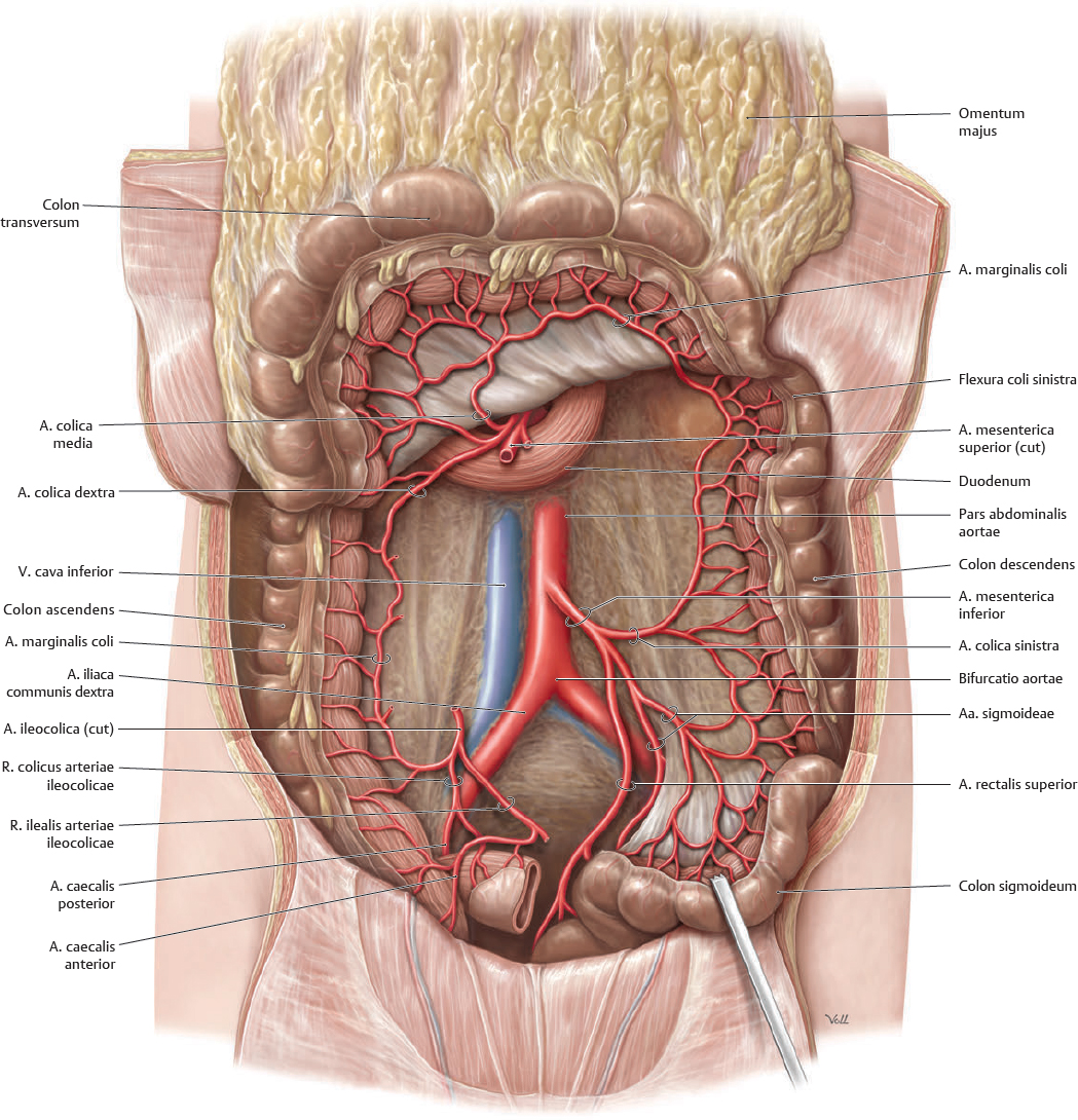
Fig. 16.12 Arteria mesenterica inferior
Anterior view. Removed: Jejunum and ileum. Reflected: Colon transversum.
Veins of the Abdomen
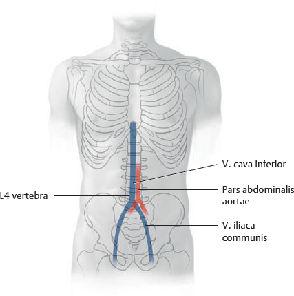
Fig. 16.13 Vena cava inferior: Location
Anterior view.
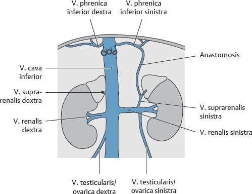
Fig. 16.14 Tributaries of the venae renales
Anterior view.
Table 16.2 Tributaries of the vena cava inferior

|

|
Vv. phrenicae inferiores (paired) |

|
Vv. hepaticae (3) |

|

|
Vv. suprarenales (the right vein is a direct tributary) |

|

|
Vv. renales (paired) |

|

|
Vv. testiculares/ovaricae (the right vein is a direct tributary) |

|

|
Vv. lumbales ascendentes (paired), not direct tributaries |

|

|
Vv. lumbales |

|

|
Vv. ilacae communes (paired) |

|
V. sacralis mediana |
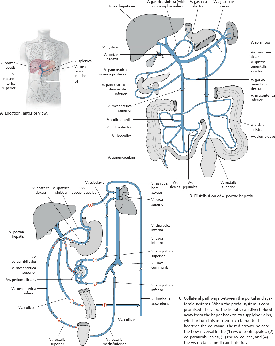
Fig. 16.15 Vena portae hepatis
The v. portae hepatis (see p. 196) drains venous blood from the abdominopelvic organs supplied by the truncus coeliacus, a. mesenterica superior, and a. mesenterica inferior.
 Clinical box 16.2
Cancer metastases
Tumors in the region drained by the v. rectalis superior may spread through the portal venous system to the capillary bed of the hepar (hepatic metastasis). Tumors drained by the vv. rectales media or inferior may metastasize to the capillary bed of the lung (pulmonary metastasis) via the v. cava inferior and the right heart.
Vena Cava Inferior & Venae Renales
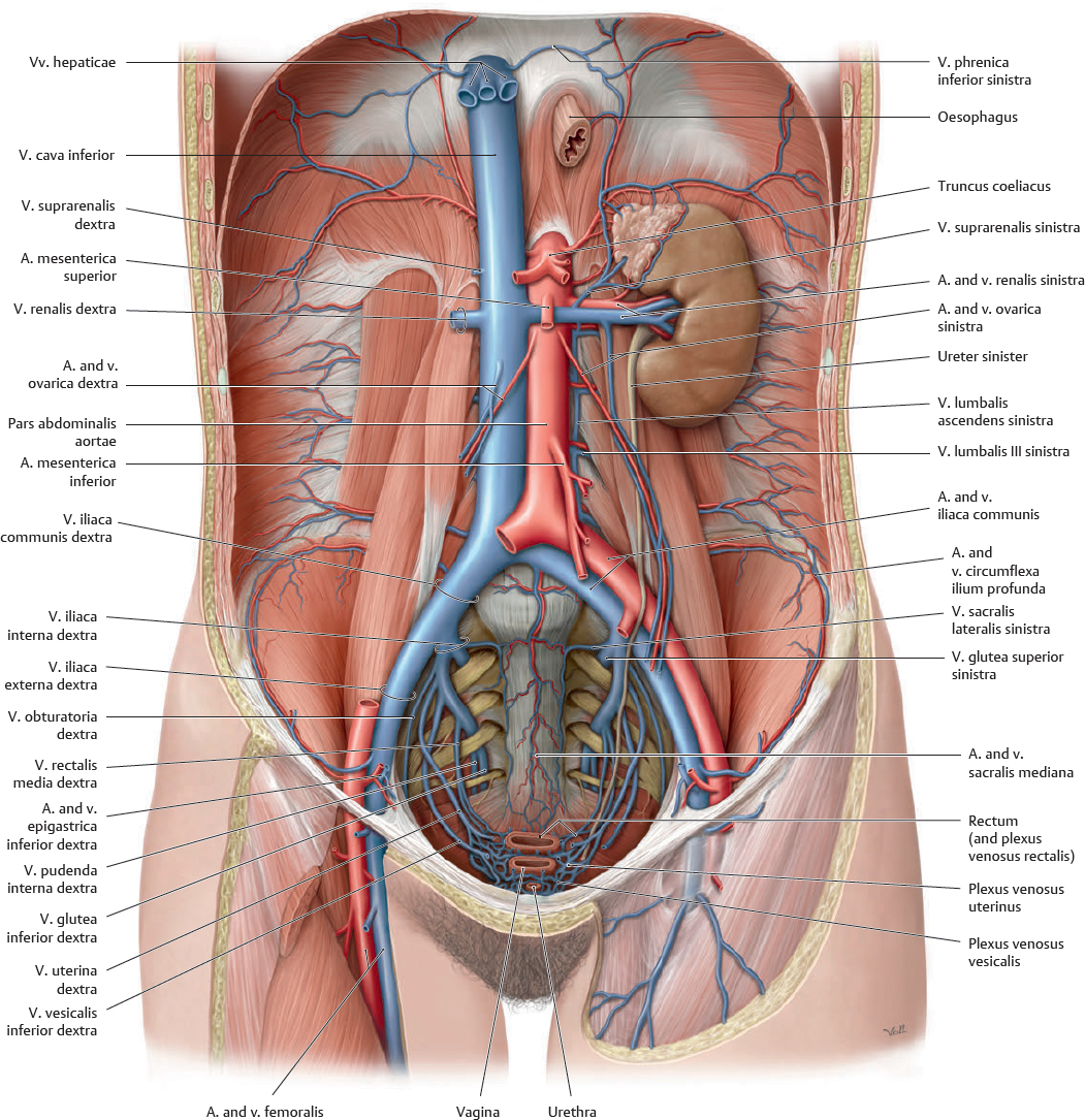
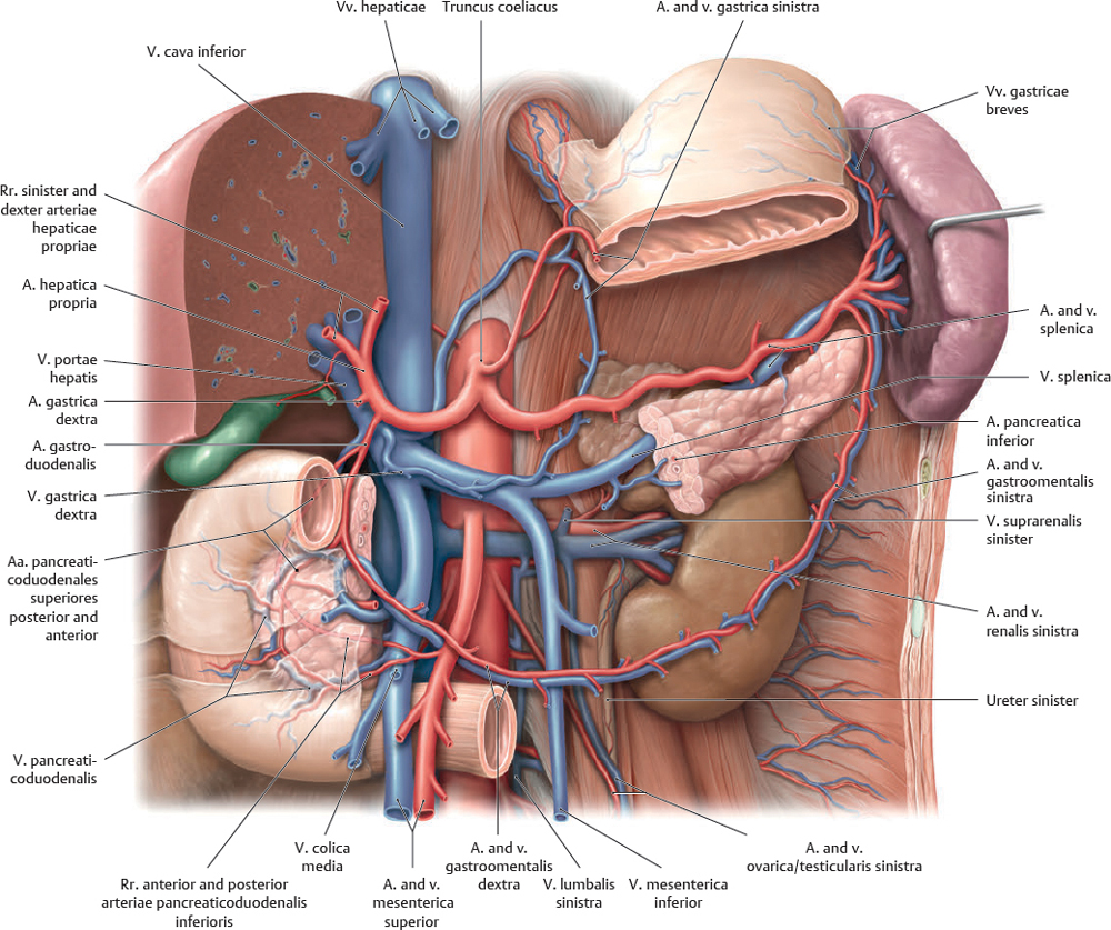
Fig. 16.16 Vena cava inferior
Anterior view of the female abdomen. Removed: All organs except the left ren and gl. suprarenalis.
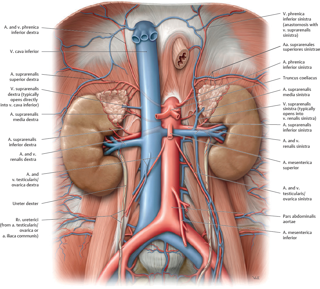
Fig. 16.17 Venae renales
Anterior view. See p. 187 for the aa. renales in isolation. Removed: All organs except renes and gll. suprarenales.
Vena Portae Hepatis
 The v. portae hepatis is typically formed by the union of the v. mesenterica superior and the v. splenica posterior to the collum pancreatis. The distribution of the v. portae hepatis is shown on p. 193.
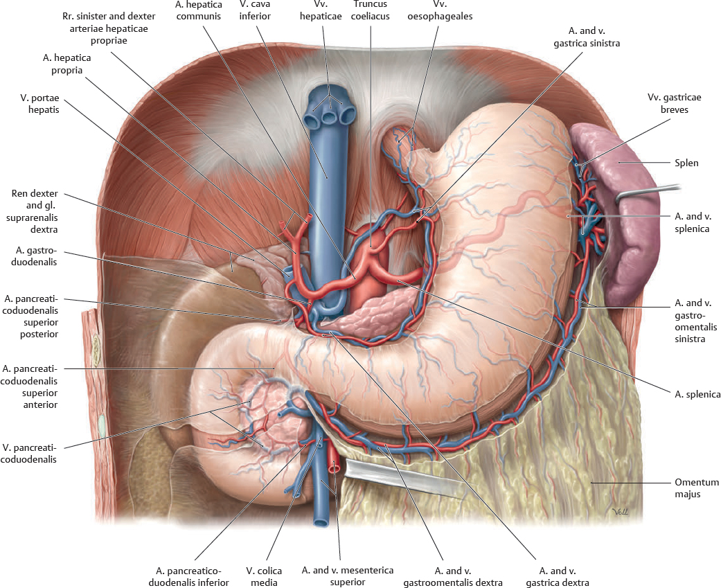
Fig. 16.18 Vena portae hepatis: Gaster and duodenum
Anterior view. Removed: Hepar, omentum minus, and peritoneum. Opened: Omentum majus.
Fig. 16.19 Vena portae hepatis: Pancreas and splen
Anterior view. Partially removed: Hepar, gaster, pancreas, and peritoneum.
Vena Mesenterica Superior & Vena Mesenterica Inferior
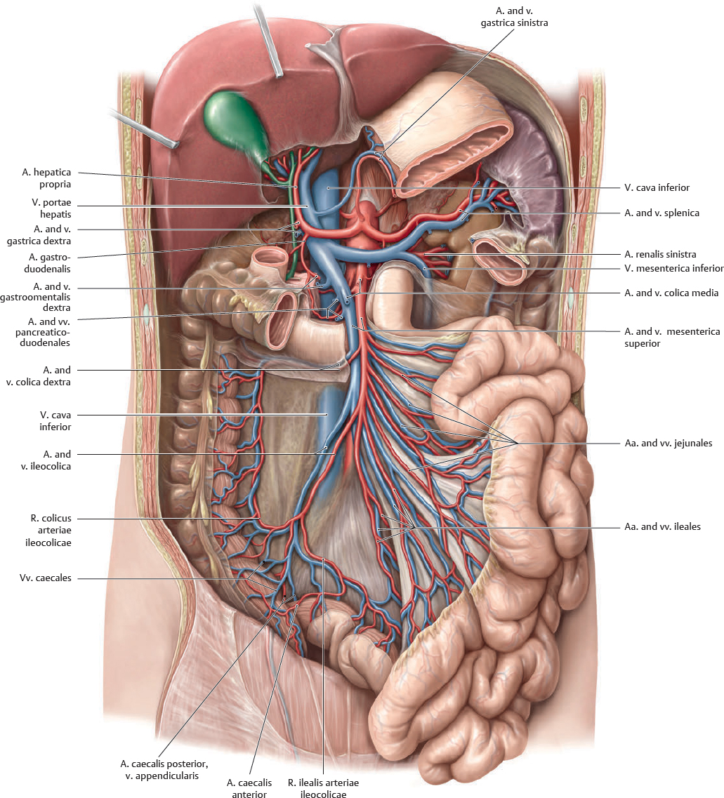
Fig. 16.20 Vena mesenterica superior
Anterior view. Partially removed: Gaster, duodenum, and peritoneum. Removed: Pancreas, omentum majus, and colon transversum. Reflected: Hepar and vesica biliaris. Displaced: Intestinum tenue.
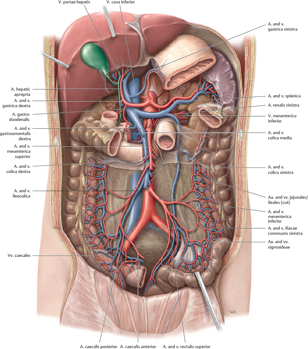
Fig. 16.21 Vena mesenterica inferior
Anterior view. Partially removed: Gaster, duodenum, and peritoneum. Removed: Pancreas, omentum majus, colon transversum, and intestinum tenue. Reflected: Hepar and vesica biliaris.
Lymphatics of the Abdominal Organs
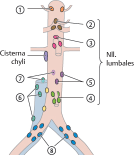
Fig. 16.22 Lymphatic drainage of the internal organs
See Table 16.3 for numbering. Lymph drainage from the abdomen, pelvis, and lower limb ultimately passes through the nll. lumbales. The nll. lumbales consist of the right nll. cavales laterales and left nll. aortici laterales, the nll. precavales and preaortici, and the nll. retrocavales and retroaortici. Efferent vasa lymphatica from the nll. cavales and aortici laterales and the nll. retrocavales and retroaortici form the trunci lumbales and those from the nll. precavales and preaortici form the trunci intestinales, respectively. The trunci lumbales and intestinales terminate in the cisterna chyli.
Table 16.3 Nodi lymphoidei of the abdomen
 Nll. phrenici inferiores Nll. phrenici inferiores
|
Nll. lumbales |
Nll. precavales/preaortici |
 Nll. coeliaci Nll. coeliaci
|
 Nll. mesenterici superiores Nll. mesenterici superiores
|
 Nll. mesenterici inferiores Nll. mesenterici inferiores
|
 Nll. aortici laterales Nll. aortici laterales
|
 Nll. cavales laterales Nll. cavales laterales
|
 Nll. retrocavales/retroaortici Nll. retrocavales/retroaortici
|
 Nll. iliaci communes Nll. iliaci communes
|
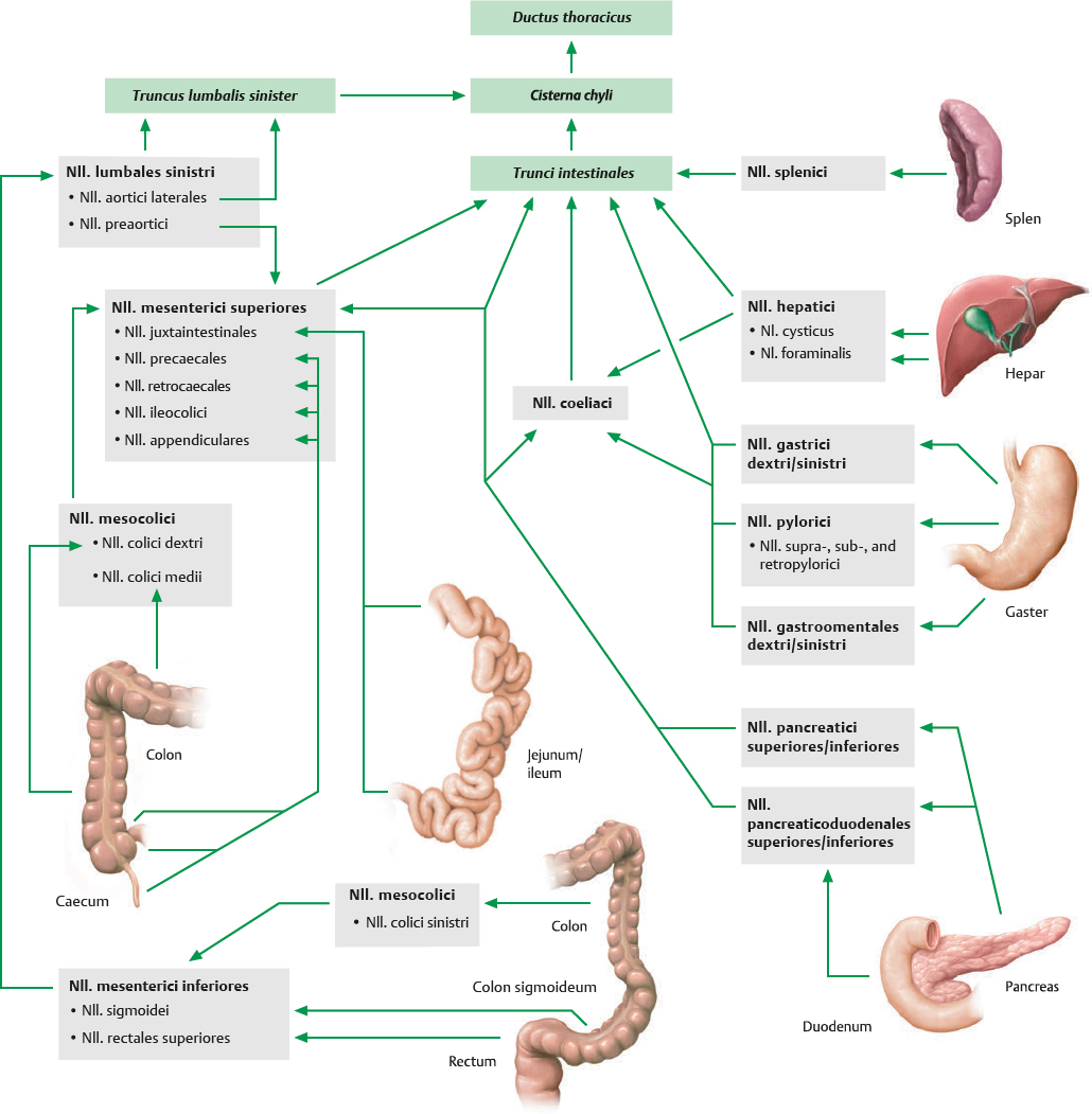
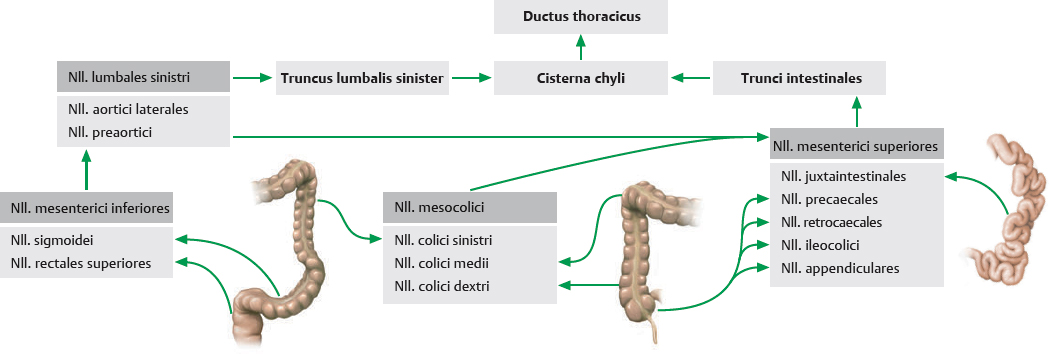
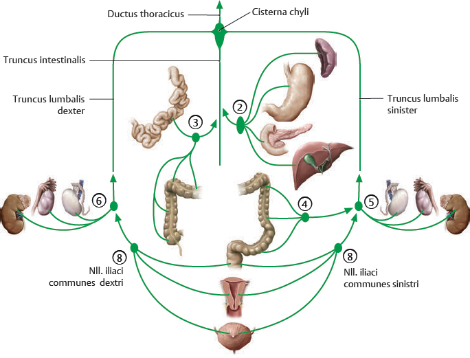
Fig. 16.23 Principal lymphatic pathways draining the digestive organs and splen
Lymph from the splen and most digestive organs drains directly from regional nll. or through intervening collecting nll. to the trunci intestinales, except for the cola descendens and sigmoideum and the upper part of the rectum, which are drained by the left truncus lumbalis.
The three large collecting nodes are:
• Nll. coeliaci collect lymph from the gaster, duodenum, pancreas, splen, and hepar. Topographically and at dissection they are often indistinguishable from the regional nll. of the nearby
upper abdominal organs.
• Nll. mesenterici superiores collect lymph from the jejunum, ileum, cola ascendens and transversum.
• Nll. mesenterici inferiores collect lymph from the cola descendens and sigmoideum and rectum.
These nodes drain principally through the trunci intestinales to the cisterna chyli, but there is an accessory drainage route by way of the nll. lumbales sinistri. Lymph from the pelvis also drains up into the nll. mesenterici inferiores and aortici/cavales laterales. A complete drainage pathway for lymph from the pelvis can be found on p. 271.
Nodi Lymphoidei of the Posterior Abdominal Wall
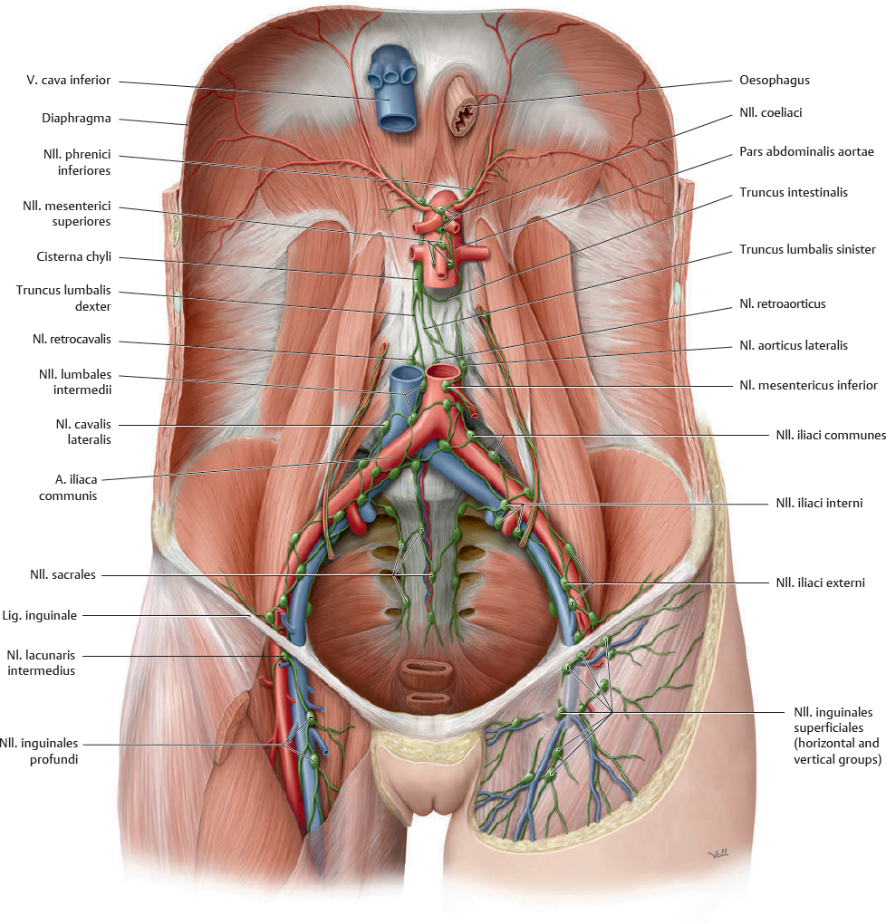
 Nodi lymphoidei in the abdomen and pelvis may be classified as either parietal or visceral. The majority of the nll. parietales are located on the posterior abdominal wall.
Fig. 16.24 Nodi lymphoidei parietales in the abdomen and pelvis
Anterior view. Removed: All visceral structures except vessels.
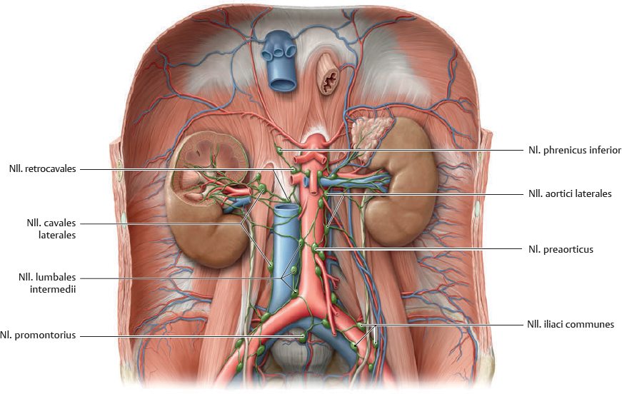
Fig. 16.25 Nodi lymphoidei of the renes, ureteres, and glandulae suprarenales
Anterior view.
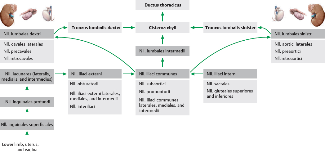
Fig. 16.26 Lymphatic drainage of the renes and gonadae (with pelvic organs)
Nodi Lymphoidei of the Supracolic Organs
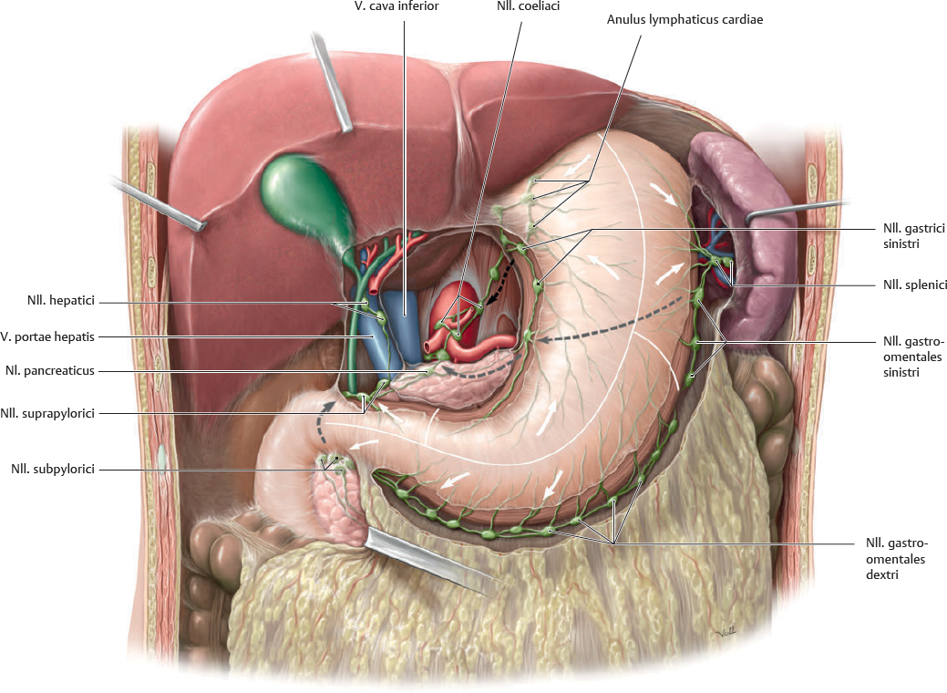
Fig. 16.27 Nodi lymphoidei of the gaster and hepar
Anterior view. Removed: Omentum minus. Opened: Omentum majus. Arrows show direction of lymphatic drainage.
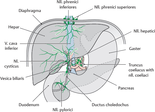
Fig. 16.28 Lymphatic drainage of the hepar and biliary tract
Anterior view. In the region of the hepar, the major lymph-producing organ, the important pathways are:
• Hepar and intrahepatic ductus biliferi: Most lymph drains inferiorly through the nll. hepatici to the nll. coeliaci and then to the truncus intestinalis and cisterna chyli, but it may take a more direct route bypassing the nll. coeliaci. A small amount drains cranially through the nll. phrenici inferiores to the truncus lumbalis. It also can drain through the diaphragma to the nll. phrenici superiores and on to the truncus bronchomediastinalis.
• Vesica biliaris: Lymph drains initially to the nl. cysticus, then follows one of the pathways described above.
• Ductus choledochus: Lymph drains through the nll. pylorici (supra-, sub-, and retropylorici) and the nl. foraminalis to the nll. coeliaci, then to the truncus intestinalis.
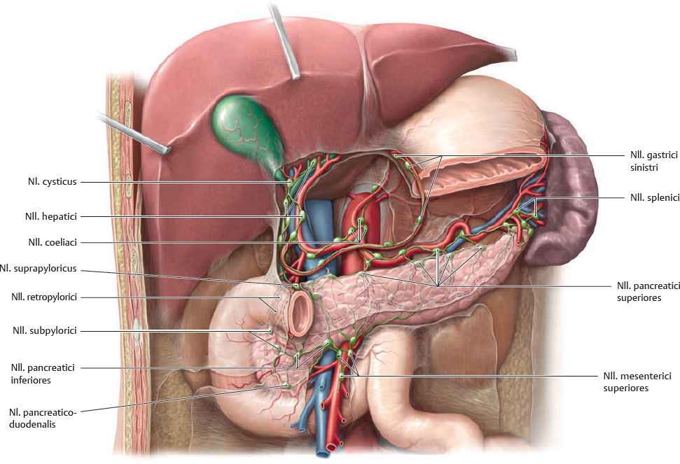
Fig. 16.29 Nodi lymphoidei of the splen, pancreas, and duodenum
Anterior view. Removed: Gaster and colon.
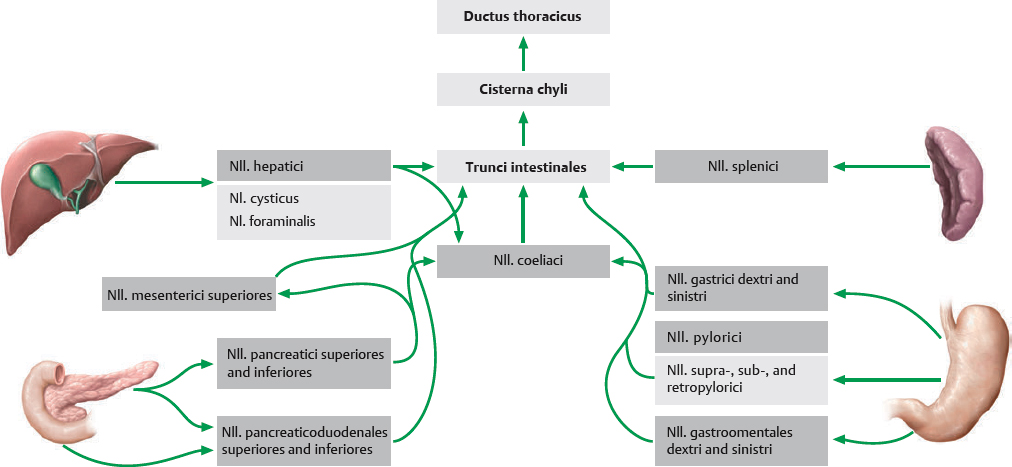
Fig. 16.30 Lymphatic drainage of the gaster, hepar, splen, pancreas, and duodenum
Nodi Lymphoidei of the Infracolic Organs
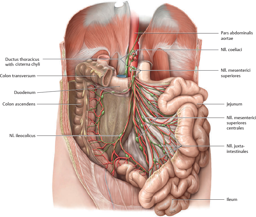
Fig. 16.31 Nodi lymphoidei of the jejunum and ileum
Anterior view. Removed: Gaster, hepar, pancreas, and colon.
Fig. 16.32 Lymphatic drainage of the intestina
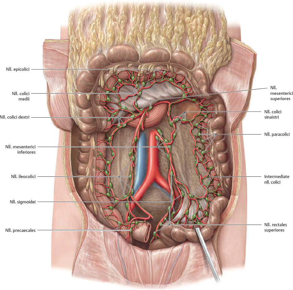
Fig. 16.33 Nodi lymphoidei of the intestinum crassum
Anterior view. Reflected: Colon transversum and omentum majus.
Nerves of the Abdominal Wall
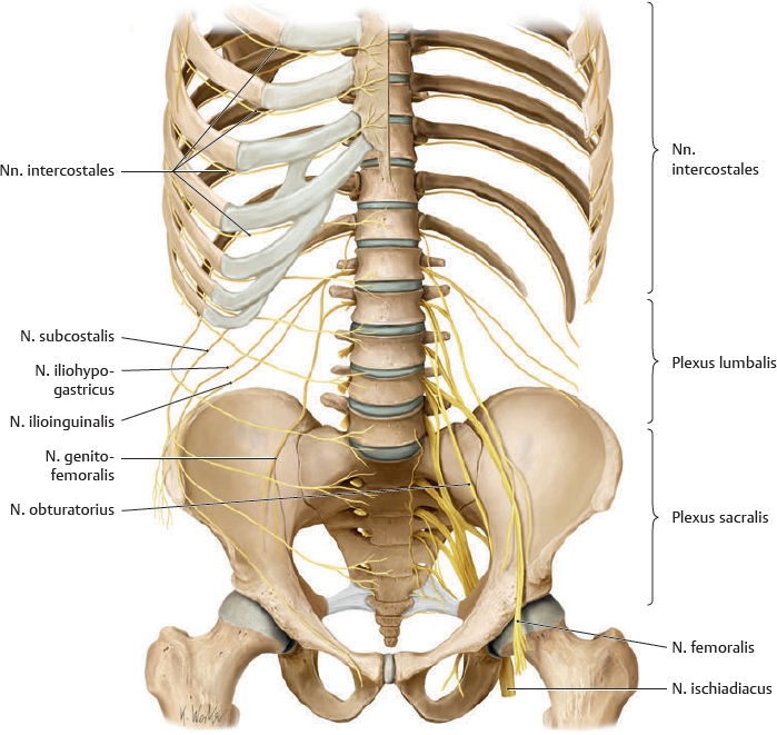
Fig. 16.34 Somatic nerves of the abdomen and pelvis
Anterior view.
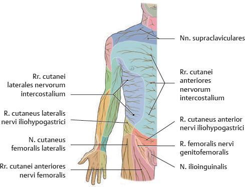
Fig. 16.35 Cutaneous innervation of the anterior trunk
Anterior view.
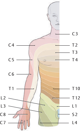
Fig. 16.36 Dermatomes of the anterior trunk
Anterior view.
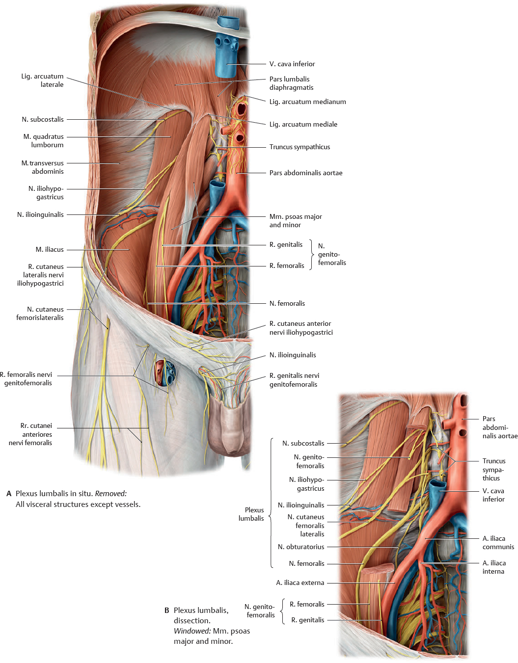
Fig. 16.37 Nerves of the plexus lumbalis
Anterior view.
Autonomic Innervation: Overview
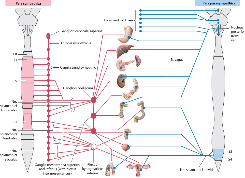
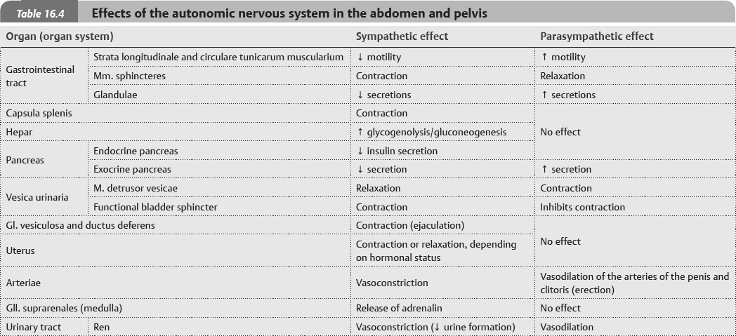
Fig. 16.38 Sympathetic and parasympathetic nervous systems in the abdomen and pelvis
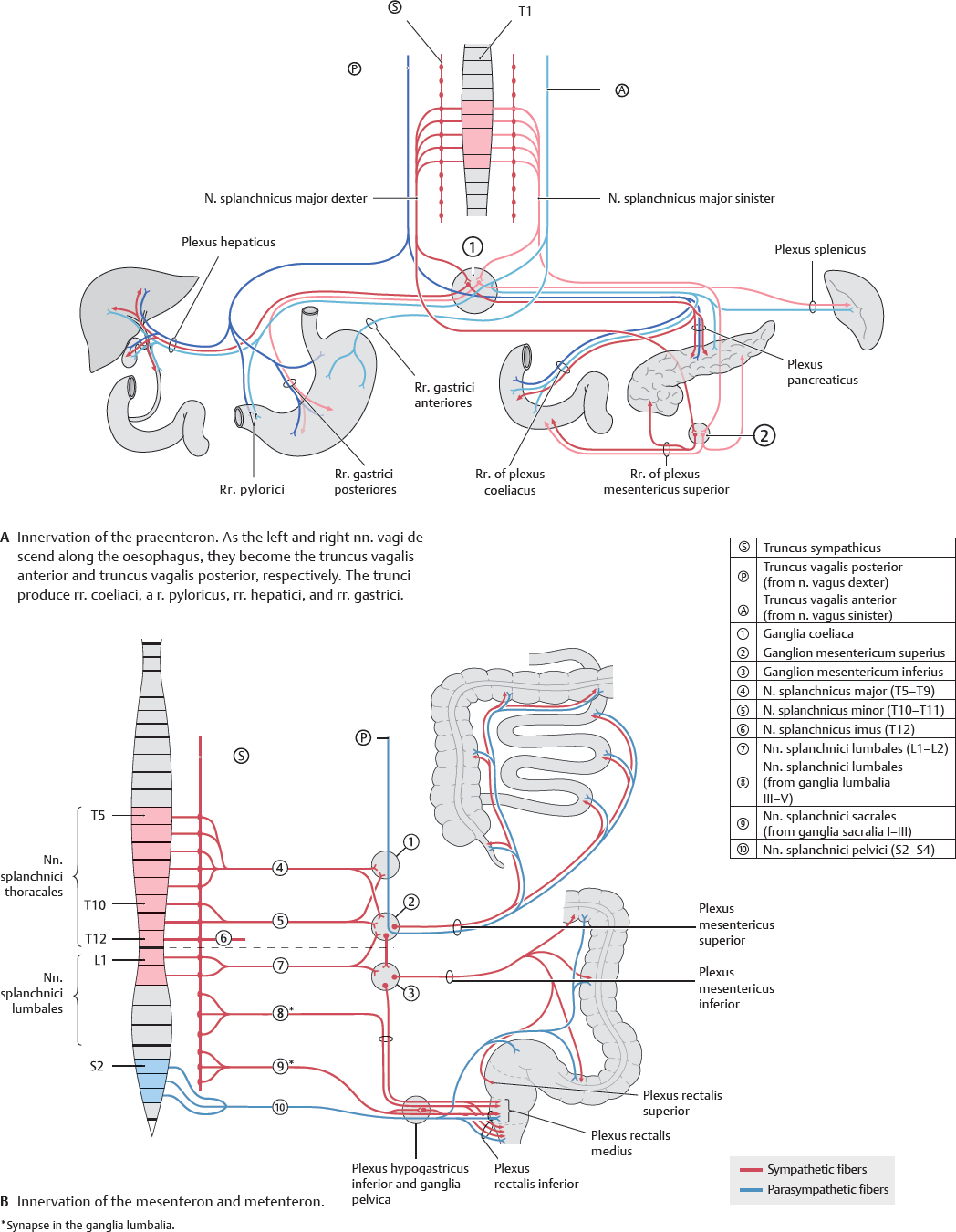
Fig. 16.39 Autonomic innervation of the intraperitoneal organs
Autonomic Plexuses
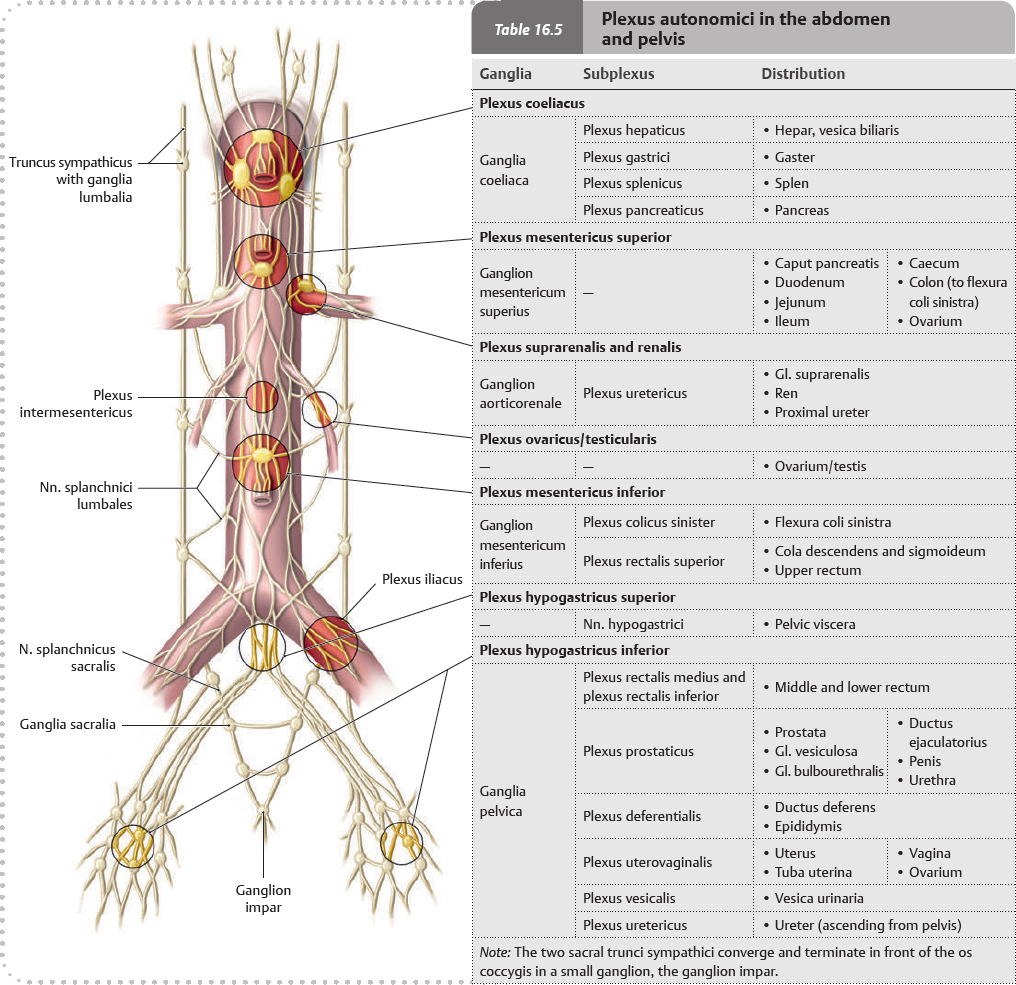
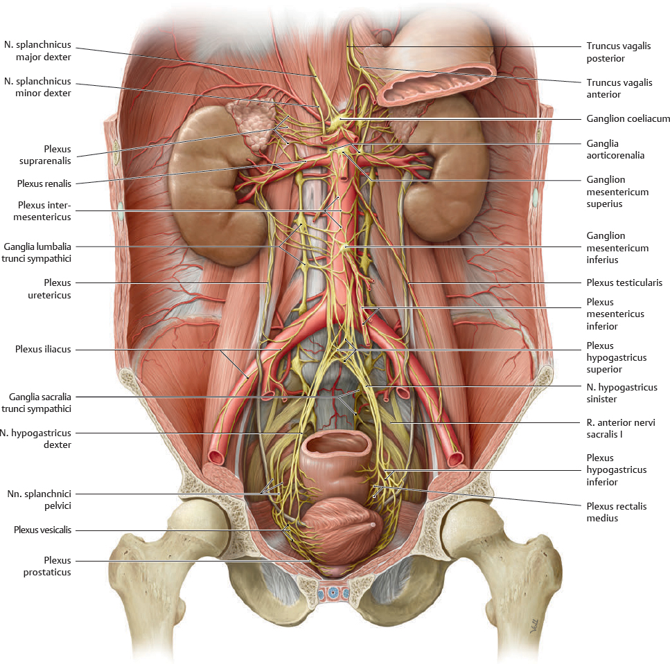
Fig. 16.40 Autonomic plexuses in the abdomen and pelvis
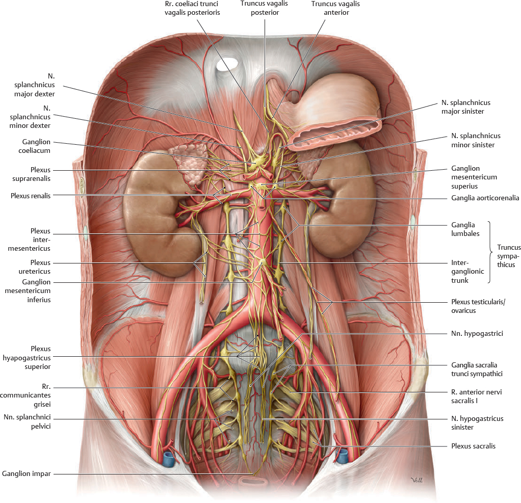
Anterior view of the male abdomen and pelvis. Removed: Peritoneum, majority of the gaster, and all other abdominal organs except renes and gll. suprarenales.
Innervation of the Abdominal Organs
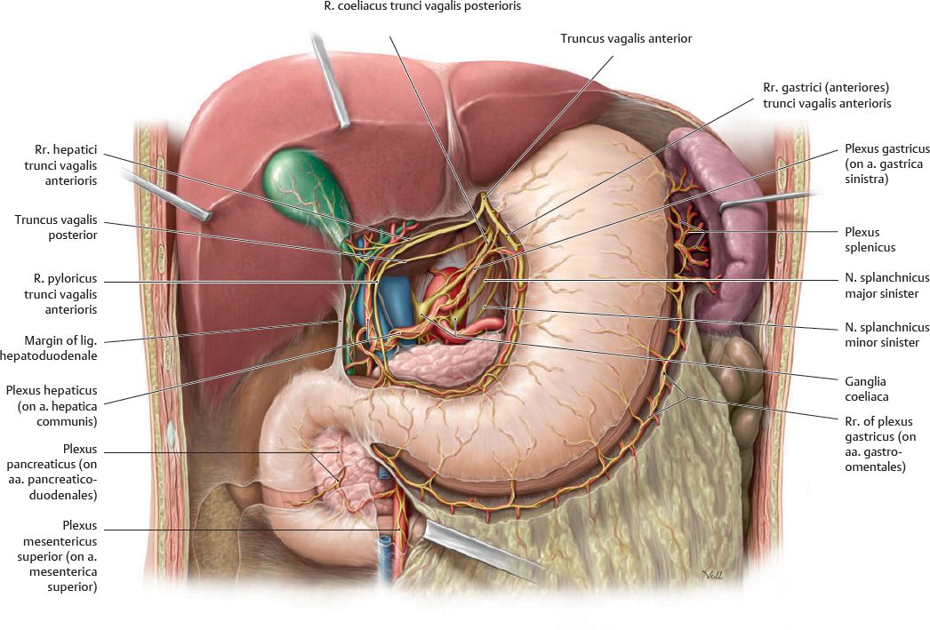
Fig. 16.41 Innervation of the anterior abdominal organs
Anterior view. Removed: Omentum minus, colon ascendens, and parts of the colon transversum. Opened: Bursa omentalis. The trunci vagales anterior and posterior produce a r. coeliacus, r. hepaticus, and r. pyloricus, and rr. gastrici anteriores and posteriores. See p. 210 for schematic.
Fig. 16.42 Innervation of the urinary organs
Anterior view of the male abdomen and pelvis. Removed: Peritoneum, majority of gaster, and abdominal organs except renes, gll. suprarenales, and vesica urinaria. See p. 276 for schematic.
Innervation of the Intestines
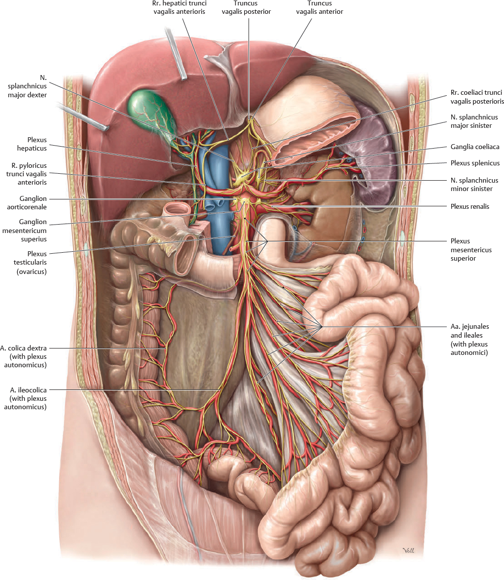
Fig. 16.43 Innervation of the small intestine (intestinum tenue)
Anterior view. Partially removed: Gaster, pancreas, and colon transversum (distal part). See p. 210 for schematic.
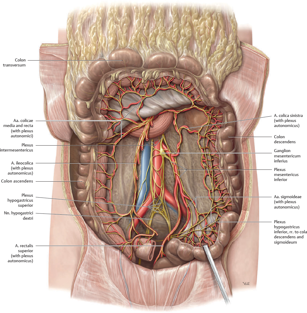
Fig. 16.44 Innervation of the large intestine (intestinum crassum)
Anterior view. Removed: Intestinum tenue. Reflected: Cola transversum and sigmoideum. See p. 210 for schematic.

 Clinical box 16.1
 The distribution of the truncus coeliacus is shown on


 Nll. phrenici inferiores
 Nll. mesenterici superiores
 Nll. mesenterici inferiores
 Nll. aortici laterales
 Nll. cavales laterales
 Nll. retrocavales/retroaortici
 Nll. iliaci communes