 Chapter 22, ‘Management: treatment’, pp. 284–285.
Skin conditions are extremely common and may occur at any stage of HIV infection. Appearance of certain skin conditions should alert physicians to possibility of undiagnosed HIV infection. Pre-existing skin conditions may worsen after its acquisition. Immunodeficiency is associated with atypical presentations, severe manifestations, and a poor response to treatment. In general, improved immunity with ART resolves or improves them.
Culture and biopsy are important in diagnosis. Particularly in patients with significant immunosuppression specimens should be sent for histology, and separately for fungal, bacterial, and TB culture. Where indicated, viral PCR may be needed.
Skin rash occurs in up to 70% cases. It is often part of an infectious mononucleosis-like illness. Typically, symmetrical and non-itchy, extending over the trunk and upper limbs, and macular or maculopapular in appearance, although it can be vesicular, pustular, or urticarial. May be associated with oropharyngeal and genital ulceration. Resolves spontaneously within 1–2 weeks.
1° and recurrent HSV present with genital and orofacial clusters of vesicles that ulcerate, crust, and heal within 2–3 weeks during the early stages of HIV infection. With advanced disease, ulcers become atypical or chronic and may coalesce to form large painful crusted lesions commonly seen peri-anally, but may involve the peri-oral and rarely the peri-ungual region. Though dissemination is rare, lesions may be auto-inoculated to distant sites. HSV infection rarely presents with necrotizing folliculitis (difficult to diagnose without biopsy).
A swab sent for HSV/PCR testing of skin lesions is very sensitive and specific. Biopsy and histological examination may be necessary for atypical presentations, demonstrating multinucleated giant epithelial cells, and immunohistochemistry can stain for HSV.
Chapter 22, ‘Management: treatment’, pp. 284–285.
Those with no previous exposure to VZV develop chickenpox, which may be severe and associated with visceral involvement.
Most adults have been infected by VZV and so present with shingles. Vesicular eruption is normally preceded by tingling and a burning sensation. In the immunocompromised affects multiple dermatomes, commonly the thoracic and the trigeminal nerves. In advanced HIV, disease tends to have painful bullous haemorrhagic necrotic lesions that may persist for several weeks and heal with severe scarring. Recurrences and dissemination  . Disseminated disease is characterized by dermatomal and non-dermatomal eruptions. Rarely, VZV presents with chronic widespread ulcers or hyperkeratotic lesions.
Clinical diagnosis is usually accurate in typical dermatomal involvement, but skin biopsy is required for atypical, chronic ulcerative, and hyper-keratotic lesions.
Prompt treatment with high-dose aciclovir  risk of dissemination and shortens its course. Clinical presentation and degree of immune deficiency determine mode and length of therapy, which is usually continued until lesions start to crust.
• IV aciclovir (10 mg/kg body weight tid) for disseminated infection, CD4 count <200 cells/µL, or with involvement of the ophthalmic division of the trigeminal nerve. May be replaced with oral valaciclovir once lesions start to crust.
• Famciclovir (500 mg tid, usually for 10 days) and valaciclovir (1 g tid, usually for 7 days) have better bio-availability than aciclovir.
• Oral aciclovir (800 mg five times daily) may be given to those with limited disease and preserved immune function.
Skin care with bathing with water and mild soap is helpful. Analgesics for pain control.
Caused by a molluscum contagiosum virus and when widespread is a marker of advanced HIV disease. Lesion is typically a flesh-coloured, 2–3 mm domed, umbilicated papule, with a faint whitish core. Those with relatively preserved immune function may have mollusca in the groin, which may be chronic. With advanced HIV disease, lesions may reach 1 cm in size and may be widespread, involving the face, trunk, eyelids, and rarely the mucous membranes (conjunctivae and lips). Mollusca are commonly seen in the beard area (related to trauma of shaving) where they are difficult to treat.
Treatment comprises cryotherapy or curettage, and is given for cosmetic reasons; no cure is available.
Widespread, resistant, and recurrent warts, which may have atypical appearances, are seen more frequently in those immunosuppressed. Facial involvement, otherwise rare, is well recognized in HIV infection.
Principles of therapy are as for those not infected by HIV. Ablative treatment is used depending on morphology, location, and number.
Staphylococcus aureus nasal carriage is common, explaining rates of infection with this organism. S. aureus skin infection are often associated with a golden yellow crusting and presents as:
• folliculitis: commonly in the hirsute areas, e.g. groin, axilla, face, and trunk; infection may involve deeper tissues, forming abscesses
• hidradenitis-like plaques: many adjacent follicles infected, forming large discoloured lesions several centimetres deep
• bullous impetigo: commonly seen on the groin and axillae as superficial vesicles or ulcers with yellow crusts
• ecthyma: an eroded or ulcerated lesion with an adherent crust covering an abscess
• scalded skin syndrome: part of systemic S. aureus infection.
Swab should be taken for culture to rule out MRSA. Superficial infection responds to standard anti-staphylococcal antibiotics (e.g. flucloxacillin 500 mg qds for 7–10 days).
Deep infection may require abscess drainage and prolonged courses of combined antibiotics, based on bacterial sensitivities. Washing the area with antiseptics helps by removing crusts and bacterial concentration.
Caused by Bartonella henselae (transmitted by flea and scratch from infected animals) and Bartonella quintana (transmitted by body louse), small Gram –ve aerobic fastidious bacilli. In addition, B. henselae causes cat scratch fever and B. quintana causes trench fever. Angiogenic lesions are most often recognized in cutaneous or SC tissues and can be difficult to differentiate from KS. B. henselae causes lesions in lymph nodes, liver (peliosis hepatitis), and spleen. B. quintana has a predilection for subcutaneous deep soft tissues and bones.
• Bacilliary angioma: friable, easy bleeding hyperpigmented papules, nodules, or plaques, but in the early stages may be purplish to bright red in colour, up to several centimetres in diameter. Lesions solitary or multiple, and widespread on the skin. Need to be differentiated from KS and pyogenic granuloma.
• Bacteraemia: presenting as pyrexia of unknown origin.
• Organ involvement: e.g. liver, spleen, brain, and lymph nodes.
• Abnormal vascular proliferation and a mixed inflammatory infiltrate on histological examination of tissues.
• Special stains: e.g. Warthin–Starry, Steiner and Steiner, required.
• PCR of tissue and blood samples.
Serological tests are not reliable in HIV infection.
Prolonged course of antibiotics, e.g. erythromycin 500 mg qds, doxycycline 100 mg bd, or azithromycin 0.5–1.0 g daily, until lesions heal.
1° Mycobacterial skin infection is rare. Skin may be involved in up to 10% of disseminated Mycobacterium avium complex infection, typically when the CD4 count is <50 cells/µL. Most frequent presentations are chronic sinuses overlying an infected lymph node (scrofuloderma) and chronic skin ulcer. Rare presentations include violaceous nodules, necrotic papules, plaques, panniculitis, and erythema nodosum.
Mycobacterial infection should be considered in any chronic non-healing skin ulcer. Diagnose by demonstration of acid-fast bacilli in smears and by culture for TB. Caseating granuloma is usually absent.
Chapter 45, ‘Tuberculosis’, pp. 531–533; Chapter 48, ‘Mycobacterium avium complex’, p. 569.
Skin infection with Candida spp. occurs in different forms, including Tinea unguium (leuconychia, nail ridging, flaking, onycholysis, and atrophy), acute paronychia (tender fluctuation of the nailbed), chronic paronychia, and intertrigo (an erosive painful erythematous rash on flexures associated with satellite pustules). Acute candidal paronychia must be differentiated from that caused by HSV infection using appropriate tests.
• Tinea unguium requires prolonged systemic antifungals (e.g. itraconazole).
• Acute paronychia and intertrigo respond well to topical antifungals (e.g. miconazole).
Very common in HIV infection. May be atypical and extensive, and may mimic inflammatory skin conditions, such as seborrhoeic dermatitis or psoriasis.
• Tinea pedis: usually presents with interdigital maceration and scaling of the soles, rarely with hyperkeratosis of the soles. Usually, associated with Tinea unguium. Caused by Trichophyton rubrum. 2° bacterial infection common and may result in cellulitis.
• Onychomycosis: results in subungual hyperkeratosis and toenail atrophy.
• Tinea cruris: symmetrical, erythematous scaling rash with central clearance on the groin, sometimes extending to buttocks and thighs. May resemble seborrhoeic dermatitis because of absence of central clearance.
• Tinea corporis: annular scaling plaques with central clearance.
• Tinea capitis: localized scaling discoid patch or generalized scaling resembling seborrhoeic dermatitis.
• Tinea faciale: differentiated from seborrhoeic dermatitis by asymmetrical distribution and well-demarcated edge.
Apart from tinea pedis, which can be treated with topical antifungals, other forms need systemic antifungals, such as terbinafine (250 mg daily for several weeks) or a triazole (e.g. fluconazole 50 mg daily).
Skin involvement occurs in up to 20% of systemic cryptococcal infection. Most common skin manifestation is a nodule or papule with central umbilication resembling Molluscum contagiosum, usually on the face. Plaques and tender SC lesions are rare. Diagnosed by skin biopsy with fungal culture and histology.
Patient should be evaluated for systemic and neurological cryptococcal infection, and managed accordingly ( Chapter 46, ‘Management’, p. 544).
Endemic to southeast Asia. Caused by Penicillium marneffei. Presents with fever, skin lesions, anaemia, lymphadenopathy, and hepatosplenomegaly. Skin lesions resemble haemorrhagic Molluscum contagiosum. Diagnosed by fungal culture of blood, bone marrow, and skin scrapings. Responds well to liposomal amphotericin followed by itraconazole, but relapses are common and long-term prophylaxis with itraconazole recommended.
Cutaneous histoplasmosis has been reported in up to 10% of patients with systemic infection, which usually occurs in endemic areas (central/eastern USA, some parts of southeast Asia, and Africa). Diagnosis by biopsy and fungal culture of papules, nodules, or ulcers. Treat with liposomal amphotericin or high-dose itraconazole followed by itraconazole maintenance.
In advanced HIV disease crusted (Norwegian) scabies may occur, characterized by widespread scaly erythematous lesions on the face and scalp, together with hyperkeratotic lesions on the hands and feet, giving the characteristic ‘breadcrumb’ appearance. Appearances can be very atypical and may require biopsy to diagnose. Highly infectious because of heavy infestation.
Patients with crusted scabies must be barrier nursed and, in addition to topical treatment (permethrin 5% or malathion 0.5%), may be given ivermectin (200 mcg/kg as a single dose). Topical steroids may be needed for eczematous nodules.
Occurs in up to 85% and may be a first indicator of HIV infection. Severity and recurrences with CD4 count. Related to infection with Pityrosporum spp., sebum production, and a genetic predisposition.
Presents as an itchy erythematous rash with a yellow greasy scale, but in severe cases, plaques and hyperkeratotic lesions occur. Usually affects paranasal area, but may involve eyebrows, post-auricular areas, and scalp. May also affect intertriginous areas and chest. Severe cases may resemble psoriasis. Facial lesions must be differentiated from lupus erythematosus and rosacea.
• Topical antifungals and steroids, e.g. miconazole and hydrocortisone.
• Scalp lesions: tar-containing shampoos, selenium sulfide, salicylic acid, and ketoconazole.
• Severe disease responds well to systemic triazoles, such as itraconazole.
A chronic disease, characterized by erythematous plaques or papules covered by silvery adherent scales. May appear for the first time or pre-existing disease may become worse with HIV infection. Several forms of psoriasis may co-exist.
• Chronic plaque psoriasis classically involves elbows, knees, and scalp, and may be associated with nail dystrophy.
• Flexural psoriasis affects axillae, groin, and intergluteal cleft; more common in advanced HIV disease.
• Guttate psoriasis presents with widespread raindrop-size lesions.
• Treatment of HIV may improve.
• Mild to moderate disease can be treated with a regular emollient plus moderately potent topical steroid, calcitriol, tar-containing ointments, or dithranol.
• Severe disease can be treated with systemic agents, e.g. methotrexate (especially in the presence of psoriatic arthritis), acitretin, and ciclosporin. Biological agents may also be considered. Closer monitoring of VL may be required.
Beware of effects of these drugs (apart from acitretin) on immune system.
Seen in up to 30% of cases. Severity and frequency as CD4 count . Xerosis (dry skin) is a common complaint and may be associated with an itchy papular scaly rash on the arms and legs. Skin may be damaged as a result of excessive scratching, resulting in excoriation, lichenification, eczematous changes, and discoloration. May be colonized with S. aureus.
Treatment includes topical steroids and emollients.
Pruritus is a common occurrence. Follicular, papular, or nodular lesions may be altered by excoriation and lichenification.
Chronic, intensely itchy, follicular rash affecting face, upper trunk, and extensor surfaces of the arms. Usually seen when CD4 count <200 cells/µL. Sterile papular, papulopustular, or urticarial papules, centred around hair follicles found. May be IgE with eosinophilia.
Responds best to phototherapy. Emollients for eczematous lesions. Anti-histamines not usually helpful. ART may improve.
Diagnosis of exclusion of other causes of pruritus. Clinical features 2° to scratching—excoriation, linear lesions, lichenified eczematous changes, and post-inflammatory pigmentation.
Regular emollients, topical steroids for eczematous changes, and anti-histamines.
Yeast overgrowth producing folliculitis through production of fatty acids and scale formation blocking follicular ostea. Presents with chronic or relapsing pruritic follicular-centred papules on the scalp, flexures, upper trunk, and face.
Oral itraconazole (preferred option) 200 mg daily for 7 days. If patient has AIDS, maintenance (200 mg od) or intermittent ‘pulse’ therapy should be considered. Other options are oral fluconazole and 2% ketoconazole cream. Scalp relapses are best treated with intermittent ketoconazole shampoo.
Chapter 54, ‘Kaposi’s sarcoma’, pp. 611–613.
Incidence since the introduction of ART. HHV-8 identified in 1994 as the causative agent. Although the skin is usual site, KS may develop in visceral organs.
Typically, KS lesions appear on the nose and hard palate, but may arise anywhere on the skin. Disease usually has an insidious course with new lesions appearing as existing ones enlarge. Rapidly aggressive disease may occur, but is rare. Initially, starts as a painless non-pruritic pink or red macule, or a papule that gradually darkens to resemble a bruise. May be surrounded by a yellow halo due to extravasated red cells (Plate 32). May be dark and difficult to recognize in black people (see Plates 34 and 35). KS lesions vary in size from a few millimetres to several centimetres (Plates 33 and 35). Extensive plaques with scaling can develop on the legs and may break down, producing local pain and oedema. Lesions on the soles may be particularly troublesome as they interfere with walking. Facial and genital KS is cosmetically unsightly. With treatment, the lesion becomes flat and the colour fades, but some pigmentation persists, even in the absence of residual tumour.

Plate 32 Cutaneous Kaposi’s sarcoma (47).

Plate 33 KS on white skin (47).
Reprinted from Warrell D., Cox T., and Firth J. (2010) Oxford Textbook of Medicine, © D A Warrell, with permission from Oxford University Press.
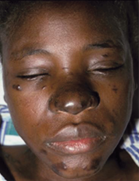
Plate 34 KS on the face (54).
Reprinted from Warrell D., Cox T., and Firth J. (2010) Oxford Textbook of Medicine, © D A Warrell with permission from Oxford University Press.
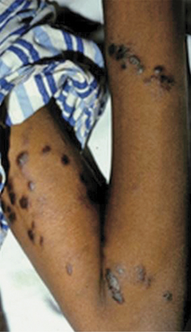
Plate 35 KS on black skin (47).
Reprinted from Warrell D., Cox T., and Firth J. (2010) Oxford Textbook of Medicine, © D A Warrell with permission from Oxford University Press.
Non-pitting oedema is commonly associated with KS, especially affecting the lower limbs. May be due to skin lymphatic involvement or 2° to vasoactive substances produced by KS. Degree of oedema occasionally disproportionate to size of KS lesions (i.e. feature of the disease itself, rather than 2° to lymph node enlargement/lymphatic obstruction).
Typical skin KS lesions are diagnosed clinically, but biopsy provides an absolute diagnosis in atypical lesions. Patients with significant mucosal involvement, fever, symptoms, or signs suggestive of other organ involvement should be evaluated by CT to look for visceral involvement or other HHV8 associated conditions. Patients with severe oedema may require CT to exclude localized obstruction or accompanying pathology.
Chapter 54, ‘Kaposi’s sarcoma’, pp. 611–613.
Treatment for skin KS is for cosmetic reasons and to alleviate symptoms.
Chapter 54, ‘Non-Hodgkin’s lymphoma’, pp. 614–616.
Extranodal cutaneous involvement occurs in up to 8% of those with B-cell non-Hodgkin’s lymphoma. Presents with an enlarging violaceous nodule or plaque, which may ulcerate.
Cutaneous T-cell lymphoma presents as a scaly patch or plaque with erythema, hypo- or hyperpigmentation, and atrophy. It may be mis-diagnosed as chronic eczema.
The skin may be involved when lymphoma (usually B-cell) involves an underlying lymph node.
Chapter 54, ‘Non-Hodgkin’s lymphoma’, pp. 614–616.
The principles of management of non-Hodgkin’s lymphoma are the same as for lymphoma elsewhere and chemotherapy is usually given.
Cutaneous drug reactions , as does the development of hypersensitivity reactions to previously tolerated drugs. Mild drug eruptions do not always necessitate the cessation of the causative drug, especially if it is the most effective agent and is given for a short time (e.g. co-trimoxazole for Pneumocystis jiroveci pneumonia). Desensitization may be possible for essential drugs. Cross-sensitivities may occur, e.g. between dapsone and co-trimoxazole.
Hypersensitivity reactions are common with certain antiretroviral drugs, such as nevirapine and abacavir, which may cause a fatal reaction. Special attention (with counselling) to patients on these treatments is important to ensure early recognition and prompt action.
• Morbilliform (erythematous maculopapular) drug eruption is common and may be associated with systemic symptoms. Most frequent causative drugs are amoxicillin and sulfonamides. Progresses in a caudocephalic direction, resolving within 3–5 days, but occasionally persisting for weeks after its withdrawal. Many viral infections cause a similar rash.
• Erythroderma occurs when a morbilliform rash becomes confluent and involves the whole body. May result in hypothermia and shock.
• Erythema multiforme, SJS, and toxic epidermal necrolysis in HIV infection. Offending drug must be stopped. If severe patients best managed in high-dependency units, where attention to electrolyte and fluid balance and skin care are of paramount importance.