Chapter 6 presented the concept of a pulse sequence and described several classes of pulse sequences. The RF and gradient pulses were applied in very precisely defined ways to uniformly affect the signal intensity from all the protons within the volume of measured tissue. Additional RF excitation pulses may be added to any of these sequences to manipulate the net magnetization of some of the tissue within the imaging volume and differentially affect its contribution to the detected signal. One approach uses frequency-selective saturation pulses, either applied in conjunction with a gradient (spatial presaturation) or in its absence (fat/water saturation, magnetization transfer suppression). Another approach uses the chemical shift frequency difference inherent in tissues to change the relative phase of the signal contribution. This is the basis for water/fat excitation using composite RF pulses and the Dixon method for fat/water suppression. In all of these cases, an increase in the minimal TR for the sequence (sequence kernel time) is required to implement the pulses. In addition, the additional RF pulse(s) increases the total RF power deposition to the patient. Limitations due to the specific rate of RF energy absorption (SAR) (see Chapter 14) may be required, particularly for spin echo-based sequences using short TR.
8.1 Spatial presaturation
8.2 Magnetization transfer suppression
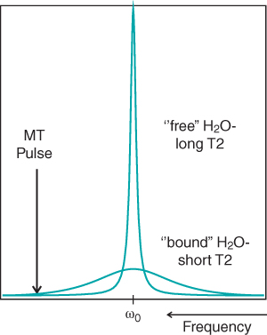
Another signal suppression technique similar in implementation to spatial presaturation is magnetization transfer suppression. A frequency-selective RF pulse is used, but in the absence of a gradient, to indirectly saturate tissue water (Figure 8.3). Water within a tissue is either mobile (freely moving) or bound (adsorbed to macromolecules). The bound fraction water protons have a very short T2 value due to the rapid dephasing they undergo. The resonance peak for these spins is very broad and normally does not contribute significantly to the measured signal. The mobile water molecules have a much longer T2 and a narrow resonance peak. These two resonances are superimposed at the same center frequency (Figure 8.4).
Figure 8.3 Standard single-echo spin echo sequence timing diagram, including a frequency- selective presaturation pulse. The saturation pulse (labeled SAT) is applied prior to the primary slice excitation pulse (labeled 90°). The RF pulse center frequency and bandwidth for the presaturation pulse are independent of these variables for the slice excitation pulses. Note the absence of the associated gradient pulse for the presaturation pulse compared to Figure 8.1.
Figure 8.4 Magnetization transfer suppression. Mobile or “free” tissue water has protons with long T2 values and produces a narrow resonance peak. Water adsorbed or “bound” to macromolecules has protons with short T2 values and produces a wide resonance peak normally not visualized in an image. Both types of water protons have the same resonant frequency. The magnetization transfer RF pulse is applied at a frequency different (off-resonance) from the water to saturate the bound water protons. Exchange between the bound and free water transfers the saturation to the free water protons, reducing signal intensity from the free water.
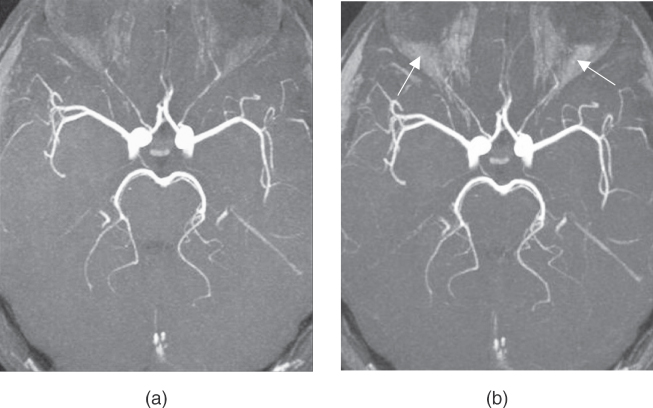
Magnetization transfer suppression is most often used to reduce a signal from normal tissue water in studies where this tissue is of little interest. Two examples illustrate this. Time-of-flight MR angiography (see Chapter 11) is a technique for visualizing blood flow within the vascular network. Suppression of the normal brain tissue water using magnetization transfer pulses enables smaller vessels to be distinguished (Figure 8.5). The other application of magnetization transfer is T1 studies following the administration of a contrast agent. T1 contrast agents shorten the T1 relaxation time for tissues where the agent is located (see Chapter 15). Comparison of images acquired before and after contrast administration enables determination of the agent dispersal within a tissue. Use of a magnetization transfer pulse during the postcontrast measurement reduces the signal from the unenhanced tissues, increasing their contrast with the enhanced tissue (Figure 8.6).
Figure 8.5 Effects of magnetization transfer in three-dimensional MR angiography. Application of MT pulse suppressed background signal from gray and white matter, enabling better visualization of blood vessels. An apparent increase in signal from suborbital fat is observed (arrows). Measurement parameters are: pulse sequence, three-dimensional refocused gradient echo, postexcitation; TR, 42 ms; TE, 7 ms; excitation angle, 25°; acquisition matrix, NPE, 192 and NRO 512 with twofold readout oversampling; FOV, RO; NSA, 1; effective slice thickness, 0.78 mm. (a) No MT pulse; (b) MT pulse.
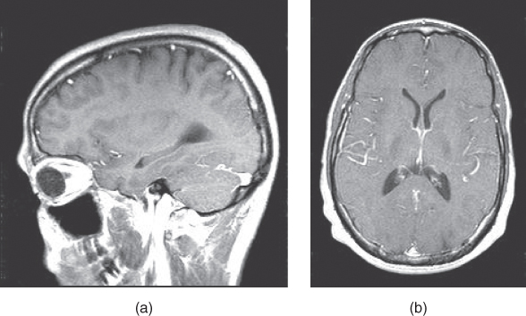
Figure 8.6 Effects of magnetization transfer in T1-weighted imaging following contrast administration. Application of MT pulse suppresses background signal from normal matter, enabling better visualization of contrast-enhanced tissues such as tumors or vascular structures. (a) No MT pulse; (b) MT pulse.
8.3 Frequency-selective saturation
While signal from either fat or water protons may be suppressed using frequency-selective presaturation, its most frequent usage is for suppression of fat. Fat saturation has two main advantages over STIR imaging (see Chapter 6) for fat suppression. It may be incorporated into virtually any type of imaging sequence. T1 fat saturation sequences may also be used with gadolinium-based T1 contrast agents since the contrast agent shortens the T1 relaxation times of only the water protons (see Chapter 15). The T1 reduction enables the enhanced tissues to generate significant signal while the fat signal remains minimal in the presence or absence of the contrast agent.
Three potential problems are inherent with frequency-selective presaturation, in addition to the problems of increased slice loop time and RF power deposition. One is that there will be magnetization transfer suppression (see above) of the water protons by the saturation pulse if fat saturation is performed. The second problem is that the saturated protons undergo T1 relaxation during the time between the saturation pulse and the imaging pulses and will contribute to the detected signal. Multiple fat saturation pulses within a slice loop may be necessary to achieve the desired signal suppression, increasing the required TR. Finally, frequency-selective presaturation is particularly sensitive to the magnetic field homogeneity. The exact resonant frequency for a fat and a water proton depend upon the magnetic field that a voxel experiences. If the homogeneity is not uniform throughout the imaging volume, the center frequency of the saturation pulse will be off-resonance for some of the spins and will not be effective in suppression (Figure 8.8b). In some cases, the water protons may be saturated rather than the fat protons. For this reason, optimization of the field homogeneity to the specific patient prior to applying a frequency-selective presaturation pulse is advisable.
 of some of the tissue within the imaging volume and differentially affect its contribution to the detected signal. One approach uses frequency-selective saturation pulses, either applied in conjunction with a gradient (spatial presaturation) or in its absence (fat/water saturation, magnetization transfer suppression). Another approach uses the chemical shift frequency difference inherent in tissues to change the relative phase of the signal contribution. This is the basis for water/fat excitation using composite RF pulses and the Dixon method for fat/water suppression. In all of these cases, an increase in the minimal TR for the sequence (sequence kernel time) is required to implement the pulses. In addition, the additional RF pulse(s) increases the total RF power deposition to the patient. Limitations due to the specific rate of RF energy absorption (SAR) (see Chapter 14) may be required, particularly for spin echo-based sequences using short TR.
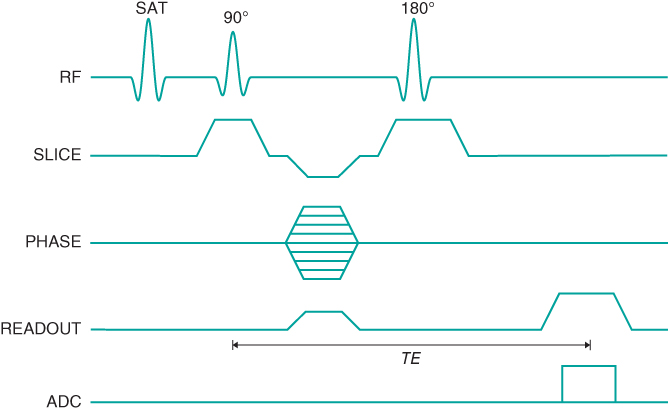
 RO; NSA, 1; effective slice thickness, 0.78 mm. (a) No MT pulse; (b) MT pulse.