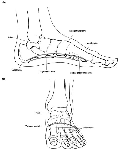
The hands and feet have the same basic structure. There are 26 bones in each foot (figure 5.1a). Starting from the distal point (the tip) there are 14 palanges; the big toe has two, a distal and a proximal, while each other toe has three: a distal, medial and proximal phalange. Then there are 5 metatarsal bones forming the foot itself and 7 tarsal bones forming the tarsus (the ankle and heel bones). The latter includes the talus, supporting the tibia and fibula (the two lower leg bones), and the calcaneum (calcaneus), forming the heel. The other bones, from medial to lateral are: the navicular or scaphoid as it is often known (which is boat-shaped, hence its name); the 3 cuneiform bones (named because of their wedged or cuneate shape), referred to as the lateral, intermediate and medial cuneiform bones; the cuboid bone (named because of its cube shape) – this is an important guide in reflexology as it denotes the waist line, and if you do not get this point correct it is easy to miss the reflexes for the two flexures (hepatic and splenic) of the large bowel.
Ligaments are tough fibrous bands of inelastic tissue, but very flexible, between the joint bones; they strengthen and support the joint, but limit movement in certain directions. There are two important ligaments in the foot: the short ligament, which extends from the calcaneum to the cuboid bone, and a longer plantar ligament, which lies nearer to the surface and supports the arch. The latter ligament is the one that practitioners must be aware of, as too much pressure on this point can be excruciating. In fact there are over 50 ligaments in each foot; these are arranged so that the sole is basically hollow.
There are three arches (figure 5.1b and c). These are, first, the two longitudinal arches. The one on the navicular side, together with the three cuneiform bones and the medial three metatarsals, forms the medial longitudinal arch. The one on the lateral side, from the cuboid notch to the distal two metatarsals, forms the lateral longitudinal arch. Secondly, the transverse arch lies across the base of all the metatarsals and is formed by the cuboid and the three cuneiform bones. This arrangement allows the lateral edge only to come into slight contact with the ground when we are walking, the calcaneus and metatarsals and the many ligaments and muscles forming the support structure of the arch.
The ball of the foot lies directly behind the heads of metatarsals. This area is very well protected with an extremely thick pad of fibrous fatty tissue to cushion and support the foot, especially when the heel is raised and locomotion is about to take place. The plantar aponeurosis is a thick mesh of collagen fibres. This not only forms a base but also allows the many muscles and tendons that are attached to it to move the bones. There are tendons to each digit and many around the ankle, all held in place by strong fibrous bands. The foot is richly served with many nerves and blood vessels, making it a vulnerable point for injury. Many people abuse their feet; it is said that each person in their lifetime could have walked over 75,000 miles, which is enough to take you around the world a few times. The pressures on the feet are greatly increased when walking or running; that is why we need this fibrous fatty pad that acts as a shock absorber. Poor posture leads to extra stresses being exerted on the ligaments and the joints, this creates discomfort and in some cases can cause the arches to become flattened or be distorted.
Tendons connect the bones with the muscles. They are made of an inelastic but flexible material. Most tendons are surrounded by a tendon sheath, a double-layered tubular sac lined with a synovial membrane (which contains fluid). The strongest and longest tendon in the body is the Achilles tendon. It commences on the calf on the posterior aspect of the leg and it inserts into the middle of the posterior compartment of the calcaneum. Two of the muscles that play a role in movement of the lower limb are the gastrocnemius and the soleus, and both of these have an insertion into the tendon of Achilles. Even though this tendon is so strong and can withstand a considerable force, it is still one of the most common sites of injury; it is supposed to be very flexible, but often in many people it becomes very tight, sore, or even swollen.
Biomechanics relates to the forces on the skeleton caused by the muscles and gravity and the resulting movements of the locomotor system. It is generally believed that many foot and leg injuries occur because of faulty alignment, although others may be hereditary.
Figure 5.1 (a) The bones of the foot. (b) The longitudinal arches. (c) The transverse arch.
The following terms are used to describe the anatomy, basic mechanics and movements of the foot, the planes of the feet and body, their motions and positions (see Glossary for those not detailed below).
• Anterior
• Posterior
• Proximal
• Distal
• Inversion
• Eversion
• Superior
• Inferior
• Abduction
• Adduction
• Sagittal
• Coronal (frontal) plane – this divides the body into front (anterior) and back (posterior). It also refers to the divisions of the foot being the hindfoot (proximal) and forefoot (distal); the movement being inversion towards the central body line or eversion away from the midline. A brief evaluation is a part of the assessment procedure.
• Transverse (horizontal) plane – this divides the body into upper-superior and lower-inferior. It refers also to the divisions of the foot being upper-dorsal, lower-plantar; the movement being adduction towards the midline, or abduction away from the midline.
• The sagittal plane – this divides the body down the middle into right and left halves. It refers to the divisions of the foot as the medial-inner aspect and lateral-outer aspect; the movement being dorsiflexion and plantarflexion.
The feet are the basic components of the kinetic chain, producing movement and acting as effective shock absorbers. They also provide the necessary information to the musculoskeletal system, so that impulses from the proprioceptors within the joints, muscles and tendons can relay the correct information to the brain to bring about co-ordinated movement. The feet are the foundations of our body, just like any building or superstructure (figure 5.1). If the underpinning is faulty, serious defects may appear later.
Spinal distortions can be caused by poor biomechanics of the feet or incorrect footwear. The wrong type of footwear, giving little or no support with inadequate cushioning, may eventually cause strain on the spine and overexert the muscles and tendons of the foot and leg. Any tautness or tension on spinal muscles can cause the cervical region to pitch forward relative to the body. If the line of gravity, which should pass through the centre of the body, is out of alignment, this can lead to faulty posture. Backs in action can cause many problems pushing, pulling, lifting, getting up and sitting down. The force of all this activity is passed down your spine to your feet. Even our heads weigh 2% of our body weight ((figure 5.2). It is of the utmost importance to assess these points at the first consultation.

Figure 5.2 How the feet support the weight of the whole skeleton
The ankle joint bears a lot of weight. It also assists in motion and acts as a shock absorber. Many soft tissue injuries over the ankle area involve the tendons and ligaments or the tarsal joint and the arch of the foot. If the foot is inflexible or stiff, it could be an injury or strain, so move it very carefully when assessing. Using reflexology directly on the area can often break down inflammation and swelling, thereby relieving the pain.
Three observation and analysis processes required for efficient analysis and diagnosis:
1. Observation as the client enters
2. Assessment through direct observation of the feet
3. Tactile awareness through touch and manipulation.
When analysing the feet we begin to build a hypothesis of the problems that may be causing an imbalance. This is not conclusive evidence, for, as holistic practitioners we need to embrace the whole person not just the feet, but we can combine all these aspects to encompass not only the physical and emotional but also the spiritual side of the individual.
Many indications must be taken into account when we use analysis or ‘external reading’ of the feet. Common foot faults need to be established first, prior to determining the nature of the disorder or imbalance. A systematic approach should be adopted so that small points will not be missed. Basic analysis of the feet is quite simple if attention is paid to detail.
Common foot disorders we need to be aware of when assessing the feet are as follows:
• Those arising from mechanical or stress defects
• Those arising from footwear faults
• Those arising from hereditary imperfections
• Those caused by infections
• Those evident indications indicative of general or systemic disorders
• Those arising from neurological disorders.
Observational skills are of paramount importance. Evaluation of the many outward superficial signs that are immediately visible may direct you to form a hypothesis regarding the person’s ailment. As practitioners, our ability to observe a person in this holistic way will provide many suggestions as to which area or system of the body may be out of balance. This evaluation should not be conclusive, because there are so many other factors. It is just a guide.
Analysis of the feet involves not only assessment of the feet when the patient is seated but also certain clinical observations. As a client walks in you should be studying the gait, noting which foot is used first to step in, as this is often the dominant side of the body. You do not have to be medically qualified to assess whether there is a normal heel strike when the person is walking. The heel should contact the ground first, followed by the forefoot. If the person has had an amputation, or has any paralysis through a stroke or poliomyelitis, this scrutiny is obviously not needed.
Abnormal structure can cause abnormal function, and the opposite is also true. Defects arise from many sources, including hereditary, or anomalies in posture and gait. The latter are being caused by behaviour patterns and other factors due to lifestyles, including obesity and stress (see chapter 3).
Variations in heel heights can cause problems. Note should be taken of the type of shoe worn. If the foot slips forward in the shoe this can cause the toes to be persistently dorsiflexed. This in turn may cause overstrain on the mechanics of the foot, such as the ankle joint, or the Achilles tendon, causing problems in the muscles of the calf. Excessive high heels may cause pressure on toes, and prolonged wearing of high heels does not allow the flexibility that the foot needs. If too high a heel is worn over a period of time, this may cause the body to pitch forward and distort the natural alignment of the body, which in turn can lead to postural abnormalities thus causing strain and tension on the abdominal organs, resulting in possible internal disorders. Faulty posture may also cause pressure to be exerted on the frontal portion of the cerebrum (brain), which may lead to depression or tension headaches.
This takes place after a full in-depth case history has been taken (see chapter 3). Make sure the patient/client is seated comfortably. Positioning and presentation are of the utmost importance (see chapter 4). This allows for a detailed examination. The therapist can observe the following suggested procedure for detailed foot observation:
1. Observe the relationship between the feet and the body. The characteristics of the feet together are comparable to those of the general body shape. For instance, a person with broad shoulders should have broad feet, a tall thin person would have long narrow feet, and so on.
2. Inspect the Achilles tendon for tightness. This tendon is involved in the lower leg muscle complex responsible for plantarflexion. Check the arches of the feet to see whether they are low or completely flat; if so, this is a sign that the spine is rigid.
3. Compare the two medial malleoli at the lower end of the tibia. A difference here indicates that one side of the hip is drawn up (see plate 6). You may also notice whether the shoulder is out of line.
4. Compare the arches of the feet, which may be different. If so, this is significant as there could be an imbalance in the musculature of the back, causing a deviation of the spine.
5. Note the suppleness of the arches of the feet. This is a good clue to how flexible the spine is.
Particular attention should be paid to the following:
• Skin elasticity – the state and quality of the skin according to age
• Skin texture – any callosities (corns or dry hard skin), blemishes or scars, lesions and moles
• Skin colour – erythema or rubor, pigmentation, pallor, cyanosis
• Skin temperature – the degree of heat or coldness: hot, warm, cold or cool (always check both feet), using the dorsal surface of the hand
• Skin humidity – sweat or moisture
• Mycotic infections – caused by a fungus (ringworm), athlete’s foot, tinea pedis
• Virus infections – plantar warts (verruca)
• Oedema – swelling or puffiness
• Dermatological disorders – eczema, contact dermatitis, allergic rashes and psoriasis.
Note that all these clinical signs may denote an imbalance within the systemic circulation. A detailed chart of clinical signs and the problems causing them is shown in Table 5.1 at the end of this section.
It is essential that the foot is scrutinized on both the dorsal and the plantar areas for any other abnormalities and clinical signs. This gives an insight into each individual’s physical attributes. Observation will provide vital clues to imbalances in certain areas, relating to organs or systems of the body.
These are imperfections or qualities passed from parent to child, although they may not be visible in all family members. All hereditary factors should be recorded. They include the following (see Glossary, for definitions): pes cavus, pes planus, pes valgus, pes varus, talipes, hammer toe, mallet toe, hallux rigidus, hallux valgus.
Note that hallux valgus and hallux rigidus can also be caused by faulty footwear, systemic or neurological disorders.
When assessing the feet, disorders of the great toe (hallux, big toe) can often be seen. Hallux valgus, the outward displacement of the great toe, is often associated with a bunion. This lateral deviation may result in discomfort on the second toe, as distortion of the great toe increases the compression on the joint. In the reflexology diagnosis it would be indicative of a problem in the corresponding area of the body, indicating shoulder or neck ailments. It could also cause an energy block throughout the whole zone or part of the zone. Any disturbance in this central zone can reflect on the whole spinal area, interrupting the flow of energy to all body parts. This in turn can affect nerve pathways and the flow of body fluids. Also, the pain and irritation can create abnormal tilting of the body, putting extra pressure on the opposite foot and hip. Even though there may be a hereditary factor here, too narrow shoes with a constricted toe-box can also cause this deformity. Additionally, if the foot is unduly broad, continuous pressure and friction over a period may cause bursitis, the inflammation of a bursa. (Bursae are small sacs of fibrous tissue lined with synovial membrane and synovial fluid, and are usually found around joints and tendons; they allow free movement.)
It is essential to give attention to the feet and lower leg to see whether there is any impaired venous drainage, which could indicate a circulatory disorder or failure of one of the valves. If there is any varicose eczema, adopt extreme care, as it is very easy to scratch and break the skin. Watches and rings should be removed for the examination, and nails should be short and well manicured to avoid damaging the patient’s skin.
Note. The therapist needs to be aware of contraindications here. For first aid relating to a varicose vein that bursts, see Appendix II.
There is an inherited tendency to varicose veins. Often you will see problems arising about 4 inches (10 cm) above the ankle and at the back and side of the calf. Look at behaviour patterns to help your assessment. Also check the dorsalis pedis point to see whether there is a good pulse. Detect the dorsalis pedis pulse (the average adult pulse rate at rest is 60–80 heart beats per minute). Press gently against the skin over the artery, using one or two fingers only. The vessel wall should feel soft to the touch. If the wall feels very hard when pressure is applied, this may be a sign of arteriosclerosis. If the rate is absent or slow, it could be indicative of some peripheral vascular disorder. If the rate is too fast, it could indicate there is an emotional disturbance caused by stress or anxiety. If unsure whether to pursue any treatment, refer the client to their medical practitioner. (See figure 5.41 ST-42.)
Nails and feet should be pink and healthy, owing to their rich nerve and blood supply. This is a good indication of the client’s health. Nail and foot disorders can be caused by several factors such as: injury, infection (bacterial, viral or mycotic), disease, pus formation, inflammation, trauma and, occasionally, nutritional deficiencies. The normal development of a nail depends on the nail’s receiving a good and adequate blood and nerve supply. Nails should also be checked to see whether they are ingrowing, involuted, cracked or chipped. Are they soft or peeling? Is the nail thickened, brittle or discoloured? Do they have transverse grooves or longitudinal striations, white spots or marks? All of these points should be noted, as they may indicate an imbalance in the corresponding area of the body (see chapter 9).
It is important to be aware of nail disorders to protect yourself and the client from cross infection.
The most common disorder is diabetes mellitus. This is due to a partial or full deficiency of the hormone insulin being produced, causing a disorder of the carbohydrate metabolism in which sugar is not oxidized to produce energy. There are two types of diabetes: non-insulin dependent and insulin dependent. The former responds to diet changes, and maybe some hypoglycaemic drug medication. Often sufferers are in the older age group.
With the latter, problems are usually more severe. Not only does the diet have to be carefully controlled with adequate carbohydrates, but also the person often needs insulin injections to maintain a normal level of carbohydrate metabolism.
Certain illnesses can cause this disorder, as well as extreme stress and anxiety. Complications the reflexologist may come into contact with are eye-related problems, so always check eye zone on the second or third toe. Kidney problems may also often arise in this disorder, and this reflex area will be very tender when we begin to apply pressure and palpation. This can be due to high blood pressure or other cardiac disorders. Tenderness may be felt all through the same zone as the eyes. The liver and pancreatic areas will also be tender. It is imperative that care is taken so that there is nothing that can scratch a patient’s skin, as loss of feeling, and conversely hypersensitivity, are common in the feet. Handle the foot very carefully and apply only the lightest of touch.
Particular problems to be aware of with this disorder include: open sores or other lesions, ulcers on the foot, vascular abnormalities, peripheral neuropathy with unexplained loss of sensations, paraesthesia and pain. These ailments make the person more at risk to infection; hence the need for extreme care because of the person’s diminished awareness of sensations.
Peripheral neuropathy may produce trophic changes (nutritional changes in the skin and other tissue), which may follow impairment of nerve supply. These include:
• Coldness
• Loss of hair
• Thinning of skin
• Abnormal sweating
• Hyperaesthesia of hands and feet
• (Tactile) exaggerated sensibility
• Impairment of sensory pathways
The client should be referred to their medical practitioner if in doubt about any signs or symptoms.
Hallux rigidus is stiffness of the joint between the great toe and the metatarsals. This can be caused by several factors, such as inflammatory joint disorders like gout or rheumatoid arthritis, previous surgery or injury to the toe through trauma, which may damage or injure the medial plantar halluces nerve, resulting in muscle weakness or sensory loss.
Hallux valgus is often due to arthritis, which starts as an inflammation. It is often followed by complications, which can cause abnormality of the joint. Hallux valgus affects the first metatarsal and the metatarsophalangeal joint, often impinging on the second toe, which then may develop into a hammer toe.
Rheumatoid arthritis is inflammation of the joints (see plate 7). The smaller joints of the feet and hands are usually involved, often affecting both limbs. The ankle joints can also be affected, with restriction of movement (see Biomechanics).
This can produce an extremely tender and painful great toe or ankle joint, and occasionally the knee joint. It is commoner in men, but women can suffer also. It is often due to the body’s inability to metabolize nitrogen-containing compounds known as purines, leading to accumulation of uric acids in and around the tissues and joints and also in the bloodstream. Over a period of time, deformity of a joint may occur. Reflexology and dietary advice are very helpful here.
The client with this disorder may experience deep pain in the heel area, which often arises from the central part of the heel at the point of attachment of the ligament structure. The pain is due to inflammation, and the problem should not be neglected, otherwise the client may develop a heel spur. Reflexology treatment to the area will improve lymphatic and venous drainage. Palpation and massage will help reduce the tensile stress at the attachment of the plantar ligament, relieving the pain and reducing the inflammation.
Disorders you may come into contact with are those that affect innervation of the limbs, either directly or indirectly controlling the hands or feet. Every muscle is controlled by a motor nerve, so if there is any sensory disturbance it can cause quite a few problems in the intrinsic muscles. One such condition is drop foot; it is due to neuritis in the anterior tibial nerve. This often causes an absence of sensation in the leg, and there is a tendency to drag the foot.
Without a good nerve supply many of the organs of the body would cease to work properly and their function would diminish. Signs of nerve degeneration include: burning sensations over the feet with itching, or pins and needles; aching in the calf area; shooting pains, or twitching or jumping in the feet or legs. Any damage or degeneration of the peripheral nerves will cause some peripheral neuropathy. Those affecting only one nerve are named mononeuropathy. The symptoms which a reflexologist may encounter are weaknesses in any of the intrinsic muscles of the foot or hand, and absence of reflexes, which may cause an unsteady gait and poor movement. There are many other causes, such as degeneration of the spinal tracts, but these do not come into the reflexologist’s remit. Dysaesthesia is a symptom of dysfunction of the sensory pathways, which may be linked to the spinal tract, causing weakness of any of the four limbs. The spinal cord is in part a receiver and an originator of many of the motor nerve impulses to and from the brain.
Reflexology has a powerful effect on the nervous system, stimulating nerve pathways. However, if there is any muscular weakness or sensory loss, the patient should be referred to their medical practitioner for investigation.
Many of the abnormalities of the feet may be caused by both neurological disorder and hereditary factors. Some abnormalities are depicted in plates 8 to 12.
Table 5.1 contains a detailed list of foot abnormalities and the disorders associated with them.
Touch is one of the vital faculties participating in the assessment technique. It not only allows you to assess and confirm a very wide range of manifestations, it also allows analysis of the tissue when palpating to distinguish whether the tissue feels solid, or whether there is evidence of deposits under the skin, or any change of texture, which may indicate an imbalance in that reflex point or zone. This technique also gives us a guide to the client’s pain threshold and will give you an insight into how to adapt your pressures when giving a full treatment session. A very marked cutaneous sensitivity could indicate nerve damage (see Neurological disorders). Always check joint movement, or any spasticity or wasting of muscles.
Just like diagnosis and professionalism, tactile skills and awareness can be achieved only with practice over a period of time. Whilst they are training, and also in the early years of practice, the reflexologist should reflect on experiences and learn from them, to develop the awareness to apply precisely the right pressure to the relevant reflex point in a systematic way.
Table 5.1 Common abnormalities of the feet, and associated disorders
This section reviews each of the body organ systems in turn and locates the foot reflex zones. Common disorders for each system are purposely not listed as most disorders are a result of an imbalance within one or more of these systems, and there can be a slight deviation from normality caused by numerous factors. The body systems do not function in isolation, but are interdependent and also rely on the finely tuned balance of each and every organ within each system. Stress is a common denominator in many disorders.
This includes all the organs involved in respiration – the nose and sinuses, mouth and throat, pharynx, larynx, trachea, bronchi, bronchioles, lungs and diaphragm, also the rib cage and the intercostal muscles. The heart and thymus are also in this anatomical area (these are covered under the circulatory and lymphatic systems respectively).
Figure 5.3 shows the organs of the respiratory system and the relevant foot zones.
The nose filters impurities in the air going to the lungs, and mucus produced by the sinuses helps to trap these. The pharynx (throat), and larynx (voice box) belong to the upper respiratory tract. The lower respiratory tract includes the trachea (windpipe), two main bronchi, smaller branching bronchioles, and alveolar sacs, where the gaseous exchange actually takes place. The lungs receive oxygen from air that we breathe in. This fresh oxygen is transferred to the blood in the tissue of the lungs. In exchange, carbon dioxide is transferred to the lung tissues, and then breathed out. Difficulties in breathing happen when this lung tissue is congested, becomes inflamed, or damaged because of pollution, smoking or infection, or if the air passages become narrowed, as in asthma. Reflexology facilitates the whole breathing process by opening up the airways. In cases of asthma, working on the adrenal glands stems any inflammation and aids the breathing process.
The foot positions for the structures of the respiratory system are shown in Table 5.2.
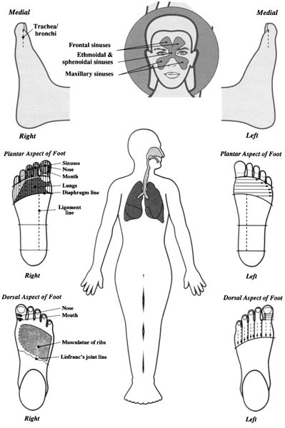
Figure 5.3 The respiratory system and its representation on the feet
Table 5.2 Foot positions for structures of the respiratory system
| Referral area | Positions on foot |
|---|---|
| Mouth/nose | Great toe medial and dorsal aspect, just below the nail bed, zone 1 |
| Sinuses | The first three toes, from the medial edge, plantar and dorsal aspect, zones 1–3 |
| Throat | Great toe medial edge and dorsal aspect, zone 1 |
| Larynx | In the web between the great toe and the second toe |
| Trachea/bronchi | In line with the spine from nose point to the middle of the proximal phalange of the great toe, zone 1 |
| Lungs | The main part of the proximal phalanges to the heads of the metatarsals, dorsal and plantar, zones 1–4 |
| Rib cage and intercostal muscles | On the dorsal aspect of the foot in the above area and down to the fifth metatarsal notch, zones 1–4 |
| Diaphragm line | Directly on the ball of the foot, zones 1–4 |
The areas of the head include the neurological centres in the brain, which includes the cerebrum (the right foot represents the right cerebral hemisphere, and the left foot the left cerebral hemisphere) and the functional areas of the cerebral cortex, the basal ganglia, the hypothalamus and thalamus, and the epicranium, which covers the muscles of the skull. Facial areas include the eyes, ears, nose (dealt with in the previous system), jaw, teeth, trigeminal nerve, tonsils and upper lymphatics. Neck-related areas and musculature are also covered here. (The brain areas are shown in figure 2.23.)
The major structures of the eye are shown in figure 5.4a. The two lids are the protective covering of the eyes. Tears spread over the surface to clean the eye. The sclera is a thick protective outer coating. The under surface of each eyelid is covered with the conjunctiva, a smooth mucous membrane. The retina is a layer of light-sensitive cells at the back of the eye. The front eyeball is filled with a watery substance called the aqueous humour and the back eyeball with a jelly-like substance called the vitreous humour. The iris is a pigmented layer.
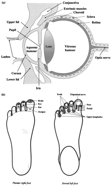
Figure 5.4 (a) A vertical section through the eye. (b) Representation on the feet.
Table 5.3 Foot positions for head-related areas
| Referral area | Position on foot |
|---|---|
| Brain area | The caps of the three first toes mainly, zones 1–5, as zones 4 and 5 merge in the head and contact the temporal area |
| Epicranial aponeurosis | The very tips of the first three toes, as near the nails as possible, zones 1–3 |
| Back of the head (occiput) | Pad of great toe, mainly zone 1 |
| Hypothalamus | See Table 5.4 |
| Cerebellum and medulla oblongata | Under the ball of the great toe, mainly zone 1 |
| Eyes | On the second/third toes, dorsal and plantar on distal phalanges, zones 2 and 3 |
| Ears | On the third/fourth toes, dorsal and plantar on distal phalanges, zones 3 and 4 |
| Facial area | Dorsal aspect of the first four toes on distal or medial phalanges, zones 1–4 |
| Nose | See Table 5.2 |
| Jaw/teeth | On the dorsal aspect of the first four toes, zones 1–4 |
| Trigeminal nerve | On the lateral and dorsal aspect of the great toe, in line with base of the distal phalange to the base of the nail bed, zone 1 |
| Tonsils | See Table 5.8 |
| Upper lymphatics | See Table 5.8 |
| Neck-related areas | Medial and lateral aspect of all the toes on the dorsum and plantar surfaces, zones 1–5 |
The structure and function of the ear are covered in detail in chapter 7.
The foot positions for head-related areas are shown in Table 5.3, and illustrated in figure 5.4b.
Many complicated functions of the body, such as body growth and development, sexual function, nutrient distribution and responses to stress, are controlled by a series of glands that produce hormones. These are known as endocrine glands; they have no ducts, and the hormones are released directly into the bloodstream and are carried to different parts of the body, where they control various aspects of the metabolism. These glands are all controlled by the ‘master gland’ – the pituitary gland at the base of the brain. The glands are scattered around the body with no anatomical link.
Glands in head-related areas include the hypothalamus, a brain area that connects the nervous system to the endocrine system by secreting releasing hormones that stimulate the pituitary gland, the pineal gland, which secretes melatonin, a hormone linked to the light-dark cycle and the body’s circadian rhythms, and the pituitary gland, which has two parts, the adenohypophysis and the neurohypophysis. Other glands are the thyroid and parathyroid, regulating calcium metabolism (the latter being four masses of tissue embedded within the thyroid), the two adrenal glands on top of the kidneys, part of the pancreas (see digestive system) and the ovaries and testes (covered in the reproductive system. The thymus has a vital role in the defence mechanisms of the body and is covered in detail in the lymphatic and immune system.
The pineal gland is thought to play a role in the light-dark cycle. The pineal gland itself is a small conical body attached by a stalk to the epithalamus on the posterior wall of the third ventricle of the brain. (Pineal is a Middle French word for ‘pine cones’.) Many theories are postulated regarding the pineal; it is thought that it may play a part in initiating the development of the gonads and the onset of puberty. The precise hormonal function is still not fully understood, but it is thought also that it may be involved with the metabolism of salt and water within the body. The pineal also contains certain neurotransmitters: noradrenaline, serotonin (thought to be the mood enhancer), histamine and some of the hormonal peptides, somatostatin, and oxytocin, a hormone secreted into the blood only by the pituitary gland that causes contraction of the uterus during labour (see Pregnancy). These hormones are produced in larger quantities elsewhere in the body. Only melatonin from the pineal is released into the bloodstream; it undergoes a nychthemeral cycle (relating to the alternation of a single night followed by day). The Pineal gland is innervated by postganglionic nerve fibres arising in the sympathetic cervical ganglia.
Figure 5.5 shows the endocrine organs and the foot positions for these areas. Table 5.4 details the foot positions for each endocrine organ.
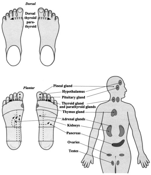
Figure 5.5 The endocrine system and its representation on the feet
There are other endocrine tissues lying in the gastrointestinal tract and producing hormones that are involved in the digestive function. The kidneys release erythropoietin, which stimulates the production of red blood cells. The placenta, an organ within the uterus, provides the embryo with the necessary nourishment and eliminates its waste; its hormonal function is the secretion of certain gonadotrophin hormones that help to maintain pregnancy.
Table 5.4 Foot positions for the endocrine glands
| Referral area | Position on foot |
|---|---|
| Hypothalamus | On the pad of the great toe, zone 1 |
| Pineal | Same area as above |
| Pituitary | On the whorl of the pad of the great toe, zone 1 |
| Thyroid/parathyroid | Mainly the base of the great toe and the second toe, dorsal and plantar aspect, zones 1–3 |
| Thymus | See Table 5.8 |
| Paired adrenals | On top of the kidneys, plantar aspect just above the pancreas, zone 1 |
| Pancreas | See Table 5.5 |
| Paired ovaries/testes | See Table 5.6 |
| Kidneys | See Table 5.6 |
As a speculative comment, since the pituitary lies in the sella turcica (Turkish saddle), the saddle-shaped portion of the sphenoid bone, which is found in the centre of the middle cranial fossa, it connects with the occipital bone; this is the reason it is thought to be only contacted by working the great toe. My conjecture is that, as it lays near the sphenoidal sinuses on this central point, it can also be contacted on the facial areas, specifically the LIV-1 point on the middle of the trigeminal pathway. This may be why this acupoint is ideal for all menstrual problems.
The digestive system includes the mouth, oesophagus, stomach and intestines, both small and large. The small intestine contains the duodenum, jejunum and ileum. The large intestine, surrounding the small intestine, consists of the caecum, vermiform appendix, and ileocaecal valve. The colon has four sections: ascending, transverse, descending and the sigmoid flexure. There are also two other flexures, the hepatic and splenic flexures. The other associated organs involved in digestion are the liver, gall bladder and pancreas.
For food to be of use to the body, it must be broken down (digested) into molecules small enough to be absorbed. Digestion of starch begins in the mouth. Food is further partly digested in the stomach. Final digestion and absorption of nutrients take place in the small intestine. In the large colon, water is removed from the contents to form the faeces, which are excreted from the rectum through the anus. The liver, gall bladder and pancreas are associated glands as their nutrients are absorbed into the body, utilized and metabolized.
The intestinal tract is another term for the gastrointestinal system. The intestines are a long tube, from the stomach to the anus, through which food and faeces pass. Many litres of fluid enter the small intestine daily. Some of this is absorbed in the small intestine, and some of this water and recycled fluids helps to hydrate the body by transferring vital electrolytes back into the system. The large intestine is involved in the formation of the faeces, and further absorption of water and essential electrolytes. Material from the small intestine enters the ileocaecal valve as liquid chyme; the ileocaecal valve is there to prevent any backflow of contents into the ileum. The ascending colon is most important as the contents have to move against gravity, and because it is a coiled tube all the flexures must be worked, the hepatic, the splenic and sigmoid flexure, so that the contents move towards the rectum. This dehydrated indigestible matter is made up from 30 per cent food waste and 70 per cent dead bacteria such as leucocytes and bile pigments. These are all squeezed along by peristalsis in the colon. The sigmoid colon is a very narrow part of the large intestine; it has no digestive function and faeces can often lie here in the rectum between the sigmoid flexure and the anal canal. The rectum is about 12cm long; if faeces remain here too long the contents can become too hard, making defecation difficult. A good general treatment, with emphasis on the pancreas area, will relieve this problem.
The hydrochloric acid secreted in the stomach has strong antiseptic qualities, necessary because of its storage capacity and its work in the gradual release of food into the duodenum; this kills off any microbes that may be present. Pepsinogen is an inactive enzyme until the hydrochloric acid activates the production of pepsin to further the breakdown of proteins. The stomach also secretes intrinsic factor that is necessary for absorption of B12, often known as the antianaemic factor because of its role in erythrocyte formation. The mucous cells that secrete the mucus are activated to release the correct quantities to act as a barrier or a protective layer. All these substances are so necessary for a balanced functioning of the digestive tract.
The liver plays a vital part in many of the processes of the body; it is our vast chemical factory. This important organ will detoxicate any noxious substances and helps in the general metabolism of fats and carbohydrates.
Figure 5.6 shows the organs of the digestive system and the reflex foot zones. Table 5.5 gives details of the foot positions.

Figure 5.6 The digestive system and its representation on the feet
Table 5.5 Foot positions for the digestive system
| Referral area | Position on foot |
|---|---|
| Mouth/tongue/teeth/salivary glands | The dorsal aspect of the first three toes, zones 1–3 |
| Throat/oesophagus | Connects the pharynx to the stomach, commencing from the base of the great toe just above the seventh cervical on the medial edge and lying behind the trachea, but anterior to the vertebral column, right down to the stomach (see note) |
| Stomach | A continuation of the oesophagus, a J-shaped area lying between this and the small intestine, zones 1–2 (right) and zones 1–3 (left) |
| Small intestine/duodenum | Starting just above the waist line by the right kidney, above the waist encircling the pancreas, zones 1–2 (right) for duodenum; zones 1–4 (both feet) for the small intestine, plantar surface |
| Large intestine | |
| Ascending colon | Working on the plantar surface, right foot only, ascending from the heel line to the waist line in a vertical strip, zones 4–5 |
| Transverse colon | Both feet following the line of the waist line, zones 1–5 |
| Descending colon | Left foot only, descending from the waist to the heel line, zones 4–5 |
| Sigmoid flexure | Just below heel line in a low shallow ‘V’ on the left foot only, zones 1–5 |
| Rectum/anus | Both feet in line with the bladder, medial aspect, zone 1 |
| Hepatic flexure | Right foot only, on the waist line and just above, zones 4–5 |
| Splenic flexure | Left foot only. As above. |
| Associated organs | |
| Liver | Occupying the main part of the area between the diaphragm line and the waist line on the right foot, zones 1–5, on the plantar aspect. The liver is basically prismatic in shape, with the left lobe extending on to the left foot, zone 1 |
| Gall bladder | Right foot on the plantar and dorsal aspect, zone 4 |
| Pancreas | The head lies in the duodenum behind the stomach, tipping up to the spleen on the plantar aspect, zones 1–2 (right), zones 1–3 (left) in line with the first and second lumbar vertebrae, just above the waist |
Note. If a person suffers from any oesophagitis, you can sometimes see a few striated lines on the right or the left foot in the area of the liver just under the diaphragm line near the medial edge. If there are any lines on the liver area it could indicate high toxicity levels or an allergy.
The zones of the reproductive system include the paired mammary glands (breasts), paired ovaries/testes, uterus/prostate, Fallopian (uterine) tubes/vas deferens. The zones of the urinary tract include the paired kidneys, the paired ureters, the urinary bladder and the urethra.
In the excretory system, the paired kidneys form and excrete urine through the ureters to the bladder, where it is stored until the bladder fills up. It then passes through a valve to the urethra, and is then expelled from the body. Waste elements in the blood are extracted in the kidneys and transformed into urine; selected substances and some water are reabsorbed. The kidneys also regulate body fluids by controlling the mineral, salt and water content in the blood.
Figure 5.7 shows the organs of the genitourinary system and the foot zones. Table 5.6 shows the details of the foot positions.
The vertebral column is a vital part of the skeletal system. As mentioned the joints and musculature allow movement. The body has a complex system of bones, joints and muscles, which allow this great variation of movement. The joints are where two or more bones meet. These are supported and strengthened by ligaments, fibrous connective tissue linking two bones together, strengthening around or inside the joints. Tendons are tough cords that connect the body of the muscle with fixed points on the bony skeleton. Some joints and tendons are lubricated by synovial fluid; this is produced by the synovial membrane surrounding the joints.
The zones of the musculoskeletal system include the spine, the neck, the musculature of the neck, the shoulder/axillary area, the shoulder point, the shoulder muscle, the arm-related area, the elbow/knee area, the hip/leg area, the musculature of the pelvis and the musculature of the buttocks. Treatment covers the neurological system, brain, spinal nerves and some peripheral nerves and the solar plexus (coeliac plexus).
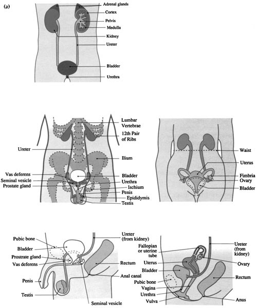
Figure 5.7 (a) The genitourinary system. (b) Representation on the feet.
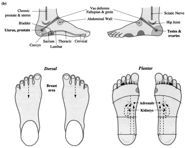
Figure 5.8 shows the musculature of the trunk and the foot zones of the muscles and skeleton (for the spinal curves see figure 2.22). Table 5.7 shows details of the foot positions.
Working the foot positions for the cervical nerves can aid most hand and arm problems, whilst working the positions for the lumbar and sacral nerves can aid most leg problems. Figure 5.9 shows the spinal innervation to the upper and lower limbs.
Note. The solar plexus lies behind the stomach; it is a network of sympathetic nerves and vagal parasympathetic fibres; it lies in zones 2–3. On the right foot, in exactly the same place, the liver receives sympathetic stimuli from an extension of the coeliac plexus and parasympathetic stimuli from the vagus nerve. This basically is one reason why we work the solar plexus reflex on both feet. As a speculative comment, this is the first point on the kidney meridian, and for all acute problems an ideal calming point for palpitations or acute problems. Which point are we working here? Is it the reflexology point? Or the nerves? Or the meridian? Does it really matter, for as previously stated this technique is empirical, and experience has shown us that this point has a wonderful way of achieving equability and restfulness in the recipient?
Table 5.6 Foot positions for the genitourinary system
| Referral area | Position on foot |
|---|---|
| Paired mammary glands (breasts female) | On the dorsal aspect (both feet), zones 1–4, with the nipple in line between zones 2 and 3 |
| Paired ovaries/testes | On the lateral aspect of the heel area (both feet) in the depression midway between the lateral malleolus and the heel area, zone 5 of the pelvic cavity |
| Uterus/prostate | On the medial aspect of the heel area on both feet in the depression midway between the medial malleolus and the heel area, zone 1 of the pelvic cavity |
| Fallopian (uterine) tubes/vas deferens | On both feet, dorsal aspect, zones 1–5. A line from the uterus/prostate point to the ovaries/testes point from the depression between each ankle across the dorsal aspect |
| Kidneys | On the plantar aspect (both feet), zones 1–2 in line with the first and second lumbar vertebrae, just straddling the waist |
| Ureters | Leading from the kidney point to the bladder, zone 1 |
| Bladder | On the medial aspect (both feet), in line with the rectum and anus point. The slightly raised area in line with the heel line, zone 1 |
| Urethra | As the bladder point |
The zones of these systems include the areas for the heart, lymphatic system, upper lymphatics and tonsils, axillary lymphatics, lymph nodes of the groin, thymus and spleen.
The cardiovascular system comprises the heart and the vast network of blood vessels, the systemic and pulmonary circulations.

Figure 5.8 Representation of the musculoskeletal system on the feet
Table 5.7 Foot positions for the musculoskeletal system
| Referral area | Position on foot |
|---|---|
| Brain | See Table 5.3 |
| Cervical spine Thoracic spine Lumbar spine Sacrum/coccyx | Inner aspect (both feet), from heel to the base of the nail bed of great toe. This area also covers the musculature of the spine as the spinae erector muscle lies above and below the line of the spine. The spinal cord is located within the vertebral column and the 31 pairs of nerves emerge along its length, zone 1 (both feet) |
| Solar plexus (coeliac plexus) | Zones 2–3 (both feet; see note) |
| Neck | Base of the first three toes, dorsal and plantar aspect, zones 1–3 (both feet) |
| Musculature of the neck | On the lateral aspect of the great toe. The medial and lateral edges of the second and third toes, zones 1–3 (both feet) |
| Shoulder/axillary area | From the diaphragm line to the tips of the fourth and fifth toes, zones 4–5 (both feet) |
| Shoulder point (see rotator cuff point, figure 5.8) | On the lateral aspect of the proximal phalange of the fifth toe. Plantar and dorsal aspect, zone 5 (both feet) |
| Shoulder muscle | On the lateral aspect of the proximal phalange of the fifth toe. Dorsal aspect, zone 5 (both feet) |
| Arm-related area | On the lateral aspect of the fifth metatarsal from the fifth metatarsal notch to the base of the little toe, zone 5 (both feet) |
| Elbow/knee | Around the fifth metatarsal notch on the lateral aspect (both feet), elbow slightly above, knee slightly below, zone 5 |
| Hip/leg | On the lateral aspect (both feet), from the heel line almost to the fifth metatarsal notch, zone 5 |
| Musculature of pelvis | Following the line of the Achilles tendon from the edge of the heel to the line of the medial malleolus, posterior to the uterus/prostate reflex, zone 1 (both feet) |
| Musculature of buttocks | Following the line of the Achilles tendon, posterior to the ovary/testes point, zone 5 (both feet) |
| Musculature of ribs | Dorsal aspect surrounding the lung area, zones 1–5 (both feet) |

Figure 5.9 Spinal nerves of the major voluntary muscles: (a) of the upper limbs; (b) of the lower limbs
It transports oxygen and nutrients around the body and is also responsible for the removal of waste products. Blood must flow through the circuit in one direction totally without being impeded or hampered in any way. In the vascular system two conditions must be maintained: (1) the competence and rhythmic regularity of the heart, (2) the right condition of all the vessels, especially the small arteries and veins. The nerve supply to these vessels is of the utmost importance as they allow the vessels to dilate and constrict.
At the centre of the circulatory system is the heart. Let’s look briefly at what work the heart has to do. It is a four-chambered very hard working double-pumping hollow muscular organ, basically a blood-filled muscular container. It acts as a double pump to drive blood around the body. One pump transports blood to the lungs to be oxygenated; the other pump has to push the newly oxygenated blood out to the rest of the body. The left ventricle receives oxygenated blood from the lungs via the left atrium and then pumps the blood, under pressure, down the aorta to the organs of the body through the arteries. From the main arteries, the blood then passes into a branching network of smaller and smaller blood vessels ending in tiny capillaries, which allow all the nutrients and oxygen in the blood to reach every cell in the body. The right ventricle receives non-oxygenated blood returning to the heart along the vena cava from the peripheral circulation via the veins and right atrium; it then pumps it at relatively low pressure into the lungs. The two sides of the heart have separate functions, but each is dependent on the other. The venous blood is kept apart from the arterial blood by the septum, a thick central muscular wall dividing each cavity. There are also four valves to prevent any backflow of blood. There is no muscle in the body so strong as the heart; each day it pumps blood through approximately 65,000 miles of blood vessels. If you are overweight then there are thousands more blood vessels and the poor hardworking heart has to work twice as hard. The heart is roughly the size of its owner’s fist.
The heart muscle contracts about 70 times a minute to pump blood around the body; to do this it needs a good supply of oxygen. It gets this from the bloodstream, not directly because it cannot obtain enough from this flow, hence it needs its own source of blood. This is supplied by its own right and left coronary arteries that arise from the aorta, and these then form branches that encircle the heart muscle. The direction of the blood flow is controlled by valves. If the arteries become too narrow because of a build-up of a fatty deposit called atheroma, the blood supply to the heart becomes restricted, or even blocked in extreme cases; this then becomes coronary heart disease, the two main disorders being angina or a more serious problem such as a heart attack.
The heart has its own intrinsic system whereby the myocardium will contract without the brain being involved, but the pace of contraction is normally controlled by fibres from the autonomic nervous system, which supply a minute area of specialized cardiac muscle located in the upper wall of the right atrium near the opening of the superior vena cava. This is the sinoatrial (SA) node, often referred to as the natural pacemaker of the body; its fibres are self-excitatory and will contract rhythmically at around 70 beats per minute. Parasympathetic fibres from the vagus nerve and sympathetic fibres from the cardiac plexus both end at this node; a fine balance is necessary at all times as it is essential that the heart contracts in a precise sequence. Extrinsic control comes from impulses originating in the cardiac centre of the medulla oblongata in the brain. These impulses from the autonomic nervous system accelerate or decrease the heart rate. Many hormones and excess stimuli of the nervous system can have an effect on the rate, either by speeding it up or slowing it down. The other area in the right atrium is the atrioventricular (AV) node (pertaining both to atrial and to ventricular chambers of the heart); this is situated slightly lower and in the middle of the heart; it receives slower impulses to contract from the SA node, through a mass of modified fibres known as the bundle of His. These originate from the AV node in front of the septum between the ventricles where they divide into two, right and left, one for each ventricle. These fibres convey nerve impulses of contraction and in response this generates tension in the muscle, causing movement in the ventricles, which then pump blood into the arteries. The pulmonary circulation is via the pulmonary artery and systemic circulation via the aorta. The nerve supply to the heart is very easily disturbed; sympathetic stimulation speeds it up, and this can be due to excitement or exercise or stress, all of which may increase cardiac activity. Cardiac activity naturally decreases while we are at rest.
Many reflexology charts show the heart in different positions, but if the feet and hands are supposed to represent the physical body then the heart’s apex must be in line with the left nipple or just below it. The heart also lies only 9 cm off the midline resting on the central tendon of the diaphragm muscle, with the left lung slightly overlapping it. The base extends up to the level of the second rib. True, there may be a false impression of referred pain to a particular area, but the reflex point is shown as the anatomy of the body.
If there is an imbalance in the heart we would be more likely to pick it up on the left foot and hand on the heart reflex, as there is more muscular bulk on the left side of the heart as it has to contract much more forcibly than the right side.
Figure 5.10 shows the heart and the relevant foot zones. Table 5.8 gives details of the foot positions.
The lymphatic circulation is part of the vascular system. The clear watery fluid called lymph passes into the blood system through the many lymph nodes and vessels situated at strategic parts of the body; these act as a filtering station, stopping foreign particles from entering the bloodstream. The lymph has to pass through many blind-ended capillaries to drain finally into two large channels: the right lymphatic ducts receive lymph that is drained from the right side of the head, thorax and the right arm; the lymph from the rest of the body drains into the thoracic duct. Lymph carries a rich supply of nutrients to every cell within the body; its other task is to remove toxins and waste. As there is no pump at its centre like the circulatory system, lymph is moved by pressure of the adjacent vessels and by muscular exercise. If it cannot flow from the tissues to the heart because of a sedentary or inactive lifestyle due to illness or injury then excess fluid may accumulate causing oedema. Reflexology is a wonderful way of encouraging the free flow of lymph; treatment helps to disperse any blockages and it also boosts the immunological system.

Figure 5.10 The cardiovascular system and its representation on the feet
The thymus, a tiny bilobed organ that plays a vital role in the immune system, produces thymosin, which stimulates the development of lymphocytes and thymopoetin, which inhibit neuromuscular transmission. Inflammation of the thymus gland (thymisitis) is thought to be responsible for myasthenia gravis, an autoimmune disorder. The spleen, another lymphatic organ, produces some types of immune cells. It is a mass of red and white pulp, controlling the quality of the blood supply through its storage of blood and iron.
Figure 5.11 shows the lymphatic system. Table 5.8 gives details of the foot positions.
Note. The general circulation is improved by reflexology, which in turn will stimulate the transportation of the necessary nutrients and hormones to the tissues, and the many leucocytes (white blood cells) to the site of infection. Oxygen supply is improved from the lungs to the tissues, which, together with the nerve and blood supply, helps the general metabolism of the muscles. The waste products of all this activity are eliminated by the excretory organs, mainly the lungs, excreting carbon dioxide and some water, the kidneys, excreting all the nitrogenous waste, principally urea, from the blood, the gastrointestinal tract, eliminating the waste products of digestion, water and bile pigments, and finally the skin, excreting carbon dioxide, some water, and a small amount of urea and salts through the sudoriferous glands.
The treatment session should commence only when the foot analysis has been completed. Each area of the foot can be worked in several ways. If there is a tender spot, go back and work it from a different angle. No one way is completely right, and each time you change direction you may pick up another congested reflex. This is therefore only a suggested order of work.
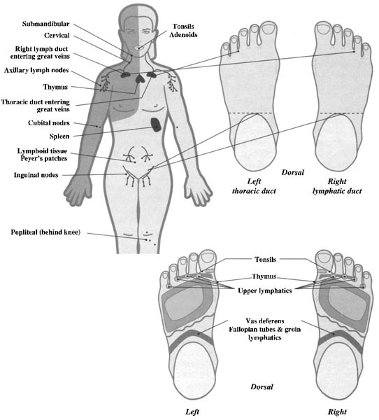
Figure 5.11 The lymphatic system and its representation on the feet
Table 5.8 Foot positions for the circulatory and lymphatic systems
| Referral area | Position on foot |
|---|---|
| Heart | In the chest area between the neck and resting on the diaphragm line. A larger area on the left foot, reaching the line of the nipple, zones 1–3. On the right foot halfway into zone 1 |
| Upper lymphatics | Webs of the toes, zones 1–5 |
| Axillary lymphatics | See Axillary area, work plantar and dorsal, zones 4–5 |
| Lymph nodes of the groin | On both feet, dorsal aspect, zones 1–5. A line from the uterus/prostate point to the ovaries/testes point from the depression between each ankle |
| Thymus | Just beneath the thyroid gland, in the fourth intercostal space just above the heart, on the very medial edge, zone 1 (both feet) |
| Spleen | On the left foot only, its base is in contact with the tail of the pancreas, zones 4–5 |
Place both thumbs on the solar plexus, as this is a good preliminary start to the treatment session. Not only does it compose you, the practitioner, but it also brings about a feeling of peacefulness to the client and it is a good way to approach the feet for the first time.

Figure 5.12 Working position for diaphragm relaxation and to commence on the lungs. This also demonstrates the support hold for working above the waist.

Figure 5.13 (a) Working all chest-related areas; separate toes accordingly, making sure you get into the base of the webs for the upper lymphatics. (b) Working the heart point on right foot. Rotate on point or palpate.
See relaxation 7 (chapter 4) and figure 5.12.
This area can be covered in vertical strips working from the medial to the lateral edge on the metatarsals and in between them; change hands and return the other way. Or this can be covered using circular friction over the whole area, or using horizontal pathways. On the medial edge there is the thymus reflex. It is important to work this area in young children or if the patient is suffering from myasthenia gravis. Change direction and repeat several times. There is a small section of the heart on this area also; again work this reflex by changing your direction. (On the left foot there is a larger area, over to zone three.)

Figure 5.14 Working the toes for all head-related areas

Figure 5.15 Working the nose area. Use firm moving pressure to release a blocked nose.
Note. Remember the area of assistance for all respiratory problems is the intestines as these aid the removal of mucus and they are also paired with the lungs in the meridian system.
Work all the toes in vertical strips. You can use both thumbs and fingers, making sure every single toe is covered on the medial edge on the plantar and the lateral edge of each toe from the medial edge to the lateral edge, and then repeat returning the other way. When you work make sure you are not in contact with the nails. When working the nose area on the dorsal aspect (see figure 5.15), make horizontal strips.
The pituitary is accessible in one of three ways. When you have completed the toes, as you come to the great toe you can push the reflex into the middle and rotate, using your left thumb on the right toe and your right thumb on the left toe. The working hand must be on top of the support hand; this ensures that the nails are not contacted. This time, working with the right thumb, pluck up and rotate; alternatively, you can work with the right thumb to right foot, pluck up and rotate – as for the eyes. Or you can apply firm unmoving pressure with your knuckle.

Figure 5.16 (a) Working the eye reflex. Remember the shape of the face; if the eyes are set wide apart this may be on the third toe. Rotate on point. (b) Pressure and rotation on the eye point, showing plucking-up method.
This is the same action as on the pituitary. You can pluck up on the reflexes and rotate using firm unmoving pressure on the dorsal aspect. Or, using both hands, press the index finger on the dorsal distal point and the thumb on the plantar distal point; rotate both together.
Note. Remember the zonal pathway and work the kidneys for eye and ear problems.
Work in horizontal strips from the great toe medial to the lateral third toe, then lateral to medial. Repeat this procedure several times. If there is a tender reflex apply circular rotation gently on the spot. Working this area also helps the eyes and ears.
This is best covered by working in vertical strips on the metatarsals and in between. Again this must be covered both ways, from medial to lateral and returning from lateral to medial. Make sure you start from the webs. If there are tender spots, work in horizontal strips, both hands working together, as in the rib cage technique (see chapter 4 figures 4.6 and 4.7), thumbs supporting the ball of the foot. On zones 4–5 you will pick up the shoulder (we will be working that area later).

Figure 5.17 (a) Working the ear point; repeat on the fourth toe. Rotate on point. (b) Pressure and rotation on the ear point, showing plucking-up method. Remember, support hand must protect the nails. Working hand is always on top of support hand.
Making a fist of the support hand provides an anchor. Now place the thumb of your working hand loosely in between the thumb and forefinger of the supporting hand, thus allowing a pincer action. Work with your index finger in tiny horizontal strips from the nail bed down to the base of the great toe and back. Work the other toes also, as in figure 5.18a.
Supporting with the flat of your right hand on the plantar surface of the foot, bring your other hand round so that you can work up the lateral side of the great toe using your left thumb. Repeat this procedure on all the toes as it is much easier to support without fear of pushing against the tiny joints of the phalanges. Change hands for the other foot, as shown in figures 5.18 and 5.19.
Making a fist of the support hand provides an anchor. Now place the thumb of your working hand loosely in between the thumb and forefinger of the supporting hand, thus allowing a pincer action. Work with your right index finger in horizontal strips from the great toe medial edge to the lateral edge of the third toe; then lateral to medial. Repeat this procedure several times. If there is a tender reflex apply circular rotation gently on the spot.

Figure 5.18 (a) Position for working the dorsal facial areas, which include the teeth, sinuses, eyes and ears. (b) Working the facial area for the teeth and mouth

Figure 5.19 Working up the lateral edge of the great toe you will contact the reflex to the trigeminal nerve.
Using the flat of the left hand, and the thumb in line exactly with the great toe, work with your right index finger up the medial edge of the toe on the trachea/bronchi reflex in line with the side of the nail.
See relaxation 10 (chapter 4). This aids and facilitates the whole breathing process.
Use relaxation 8, 9, or 12 (chapter 4). Any one, or all of these, can be utilized.
The area from the waist to the diaphragm line can be covered in slight diagonal strips working both ways from the medial to lateral edge then returning the other way; this is easier than going across in horizontal bands as there is too much loose flesh. If there is a tender spot, change direction or do circular rotations. The gall bladder lies in zones 3–4, dorsal and plantar.

Figure 5.20 Working the reflexes for the neck, thyroid/parathyroid on the dorsal aspect of the foot.
Note. You are working from the waist and above so you should be supporting from above. Watch out for the ligament line at this point as it may be very tender. Plantar flex the toes when working to safeguard against causing any undue discomfort. Remember, the nerve supply to the whole alimentary tract is balanced by the sympathetic and parasympathetic nervous systems; you may need to go back and work the brain areas.
Using your left thumb, hook out on this reflex at least three times. It lies exactly on the heel line, approximately one thumb-breadth in. You can also apply extra pressure by utilising the other thumb as well.
Note. You are now working from the waist and below so you should be supporting from underneath the heel.
Still using your left thumb, work up the lateral edge of zones 4–5 to the hepatic flexure. (See figure 5.6.)
Working with your right thumb from the medial edge to lateral edge in horizontal strips and making as many bands as you can, carry on right down to the base of the heel. Change hands and repeat the other way. Just to make sure you have contacted the hepatic flexure, go back and apply pressure on the lateral plantar side of zone 5 in line with the fifth metatarsal notch, as in figure 5.21. If there is a tender spot then change direction or do circular rotations.

Figure 5.21 Applying extra pressure on the ileocaecal value reflex
See relaxation 11 (chapter 4).
These reflexes are best worked in unison. Using the thumb of the right hand, make tiny steps on the bladder reflex. This is the slightly raised rounded area on the side of the foot in alignment with the heel line. Repeat at least three times. Then carry on up the medial edge, making sure that you do not press on the ligament line. Go right up to work on the adrenal glands point (this lies approximately two finger-breadths below the diaphragm line); rotate on this point firmly three times. Come back to the bladder and now complete a continuous circuit from the bladder up to the waist. Lift the wrist so that you can cross the waist to the kidney, making sure that you plantar flex the foot so that you do not apply undue pressure on the ligament. Once you are in zone 2 work the kidney point by rotation three times at least (see figure 5.24). This is an area that is often incorrectly worked. Normal kidney function is needed for good health. Look at figure 5.7b to see the recommended pathway for working.

Figure 5.22 (a) Working the intestines: the support hand is on the waist line and acts as a guide for the transverse colon reflex. (b) Working from the lateral to the medial edge: the support allows the skin to be relaxed and not taut.

Figure 5.23 Working the bladder point and kidney point
Note. The support hold encircles the foot in a caressing way with the left hand; the right hand is then ready for working. Take the foot out to the required position.
Apply firm unmoving pressure to the kidney reflex with the right thumb. This point should lie fractionally above the waist. The left hand rotates the foot down on to the unmoving thumb. You can also use the knuckle in sweeping downward strokes, providing it is not too tender.
This reflex can be worked with two fingers on the medial side of the heel. Work from just before the depression on it and just past it. This ensures the whole area has been covered. (See figures 5.7b for area to work and 5.31a for correct support hold.)
Figure 5.24 Pressure on the kidney reflex and rotation to aid in elimination. Right thumb is between zones 2 and 3 on the waist line.
Note. When you are working on the medial or lateral edge of the heel areas, keep the foot as straight as possible; this permits the area to be worked without the creasing of skin on the heel area hindering movement. Remember that in females there is an anatomical and physiological connection between the breasts and uterus, so the breasts will be an area of assistance.
SP-6 on the Spleen meridian shares the same point as the chronic uterus reflex; this can be used providing the person is not menstruating or pregnant, as not only is it an empirical point to promote delivery during labour, but it will also stimulate blood flow.
If there are no contraindications you can carry on from the last point up the leg for about three to six finger-breadths as near to the posterior edge of the tibia as possible; repeat this three times.
Warning! This area can be very tender. There is no need to apply great pressure. If the client is male make sure you do not drag the hairs on the legs.
This is worked exactly the same as the uterus/prostate reflex, but this time you are working on the lateral aspect of the heel. (See figure 5.7b for area to work. Change support hand.)

Figure 5.25 Working the vas deferens and Fallopian tubes and groin area
Support above with the right hand, taking the weight slightly off the heel area so that you can work with the thumb and index finger of the left hand. Start in line with the medial malleolus and work at least six finger-breadths up the Achilles tendon. Repeat several times. (See figure 5.31b.)
Support is given by cupping the heel with the fingers of both hands, and the thumbs (or index fingers) then work distal to the two malleoli up each ankle to join in the middle. Be careful not to pinch the skin. Carry on over the top with one thumb at least three times. Repeat this procedure, but this time work on the proximal side of the malleolus.
The support is from above with the right hand. Put the index finger or second finger on the prostate/uterus reflex and the thumb on the testes/ovaries reflex. Apply firm unmoving pressure as you rotate the foot with the right hand in a medial or lateral direction, as required (see relaxation 14/15, chapter 4).
This is a light hold; a grip is too strong. The web between the thumb and index finger of the left hand is placed on the above reflex, the thumb to the lateral edge. Supporting with the right hand, rotate in a medial direction, pushing up to make contact with your hand. Do this several times as it relaxes this area (see relaxation 16, chapter 4).

Figure 5.26 How to work the coccyx/sacrum on the medial edge; the support hand and working hand change over to work the lateral edge.

Figure 5.27 (a) Working all spine-related areas, including the erector spinae muscle and nerve pathways. (b) Working the spinal pathway; move the support hand up as you ascend the foot.
The aim is to cover the calcaneum almost in a ‘U’ shape. This is achieved by supporting above with the right hand as the left hand encircles the heel. Use the first three fingers starting from the midline of the heel and walk up in alternating steps to the level of the heel line of the medial edge. Change hands and repeat on the lateral aspect for the hip and pelvic area.

Figure 5.28 (a) Working the brain area. Fingers or thumbs can also be used. Work both ways, rolling the knuckle across. (b) Working the brain area using the thumbs.
The support hold encircles the foot in a caressing way with the left hand. The right hand is then ready for working; take the foot out to the required position. The spine reflex can be worked using the thumb following the line of the spine on the inner aspect of both feet, from the heel to the base of the nail bed of the great toe; this includes the cervical, thoracic, lumbar spine and sacrum/ coccyx. This area also covers the musculature of the spine as the spinae erector muscle lies above and below the line of the spine. The spinal cord is located within the vertebral column and the 31 pairs of nerves emerge along its length; if we work directly on the arch of the foot and then above and below we contact all the areas of the body. You can also work individual areas crossways, using one or two fingers. The brain area lies on the caps of the first three toes. Work this with the thumb and index finger in alternating steps over the area. Or use the knuckle in a rolling action back and forth to cover all areas. (See figure 4.10a.)
Self-help tip. If you are feeling under the weather and do not have time for a full treatment for yourself, partner or colleague, work the spine area for 5 minutes. It is a wonderful first-aid treatment.
The support is the left thumb on the base of the distal phalange and the third finger on the dorsal aspect in the same area. The second finger can then keep the toes apart. Work the lateral edge of the first three toes using the index finger or the thumb of the right hand, whichever is more comfortable.
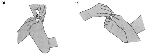
Figure 5.29 (a) Working the chronic neck area down the lateral aspect of the first three toes. (b) You can use your index finger, or the other hand.

Figure 5.30 Support for working down the spine. Use the thumb or index finger, whichever is more comfortable.
The aim is to pick up a congested spot that you may have missed as you ascended the spinal reflex. Support using the dorsum of your right hand to the plantar aspect of the right foot. Your index finger should be in line with the great toe, and your left thumb is going to work all the way down. Be sure to cup the toes as you start; place them on the lateral edge between the great toe and the second toe. If the span of your thumb is insufficient to allow you to come down easily, put the support hand under the heel and elevate the foot towards the working hand.

Figure 5.31 (a) Working the musculature; make free passes and repeat on the lateral side. (b) Squeezing technique for rectum and haemorroids.
Work the musculature of the pelvis following the line of the Achilles tendon from the edge of the heel to the line of the medial malleolus, posterior to the uterus/prostate reflex of both feet (zone 1). Then work the musculature of the buttocks following the line of the Achilles tendon, posterior to the ovary/testes point of both feet (zone 5). These reflexes lie behind the prostate/uterus/testes/ovaries respectively in the web of the heel to the last crease. An alternative is to use a squeezing technique (figure 5.31b).
See relaxation 3 (chapter 4).
Both hands are placed on the medial edge of the spinal reflex, in between the web of the thumb and the index finger. The thumbs are placed on the plantar area with the fingers lying across the dorsum. Holding the hand nearest to the ankle stationary, tweak up with the hand nearest to the toes; repeat this procedure two or three times. Then applying a stationary firm hold, gently stretch all spinal areas.
See relaxation 2 (chapter 4).
See relaxation 21 (chapter 4).
Figure 5.32 Spinal tweak and stretch
See relaxation 5 (chapter 4).
By making a fist of the support hand, the left hand knuckles to the lateral edge in a punching action; this can rotate any way and it provides an anchor. Now place the thumb of your right hand, your working hand, loosely in between the thumb and forefinger of the supporting hand, thus allowing a pincer action. If you hold it high enough this should enable you to reach over the top of the foot and work down between the third and fourth toes. As you come in line with the diaphragm line take your thumb out thus allowing you now to continue off to the outside edge in a slightly diagonal direction. Repeat this on toe 4 down the metatarsal, following the same line. Continue in between the fourth and fifth toes and then finally on the fifth toe down to the diaphragm line.
Immediately change hands; the support is with the right hand for the right foot this time. The knuckles are in line with the base of the toes, and the working hand is your left index finger. Your left thumb is placed loosely in between the thumb and forefinger of the supporting hand, thus allowing a pincer action. Work the shoulder muscle reflex three times, from the lateral edge to the third toe.
This reflex lies on the lateral aspect of the foot from the fifth metatarsal notch to the little toe. The support is the flat of your right hand, the palmar surface to the plantar area of the foot. With your hand apply slight pressure. You can move the foot into the required position encircling the foot in a caressing way; taking the foot towards the midline, work up the arm-related area at least three times.

Figure 5.33 Working the shoulder and auxillary areas
Note. For all hand problems you are working on the feet, the cross reflex. Work the area that corresponds to the problem; also the nature of the nerve pathways is such that you should also work the cervical spine area for any problems of the upper appendicular limbs. (See figure 5.9a.)
The support is from above with the right hand; this helps to expose the fifth metatarsal notch. This is a shared reflex, with the upper area serving the elbow and the lower area serving the knee. Using your index and second finger together, work above, on and below this reflex. Repeat and come back up, below, on the notch and above it, covering the area as shown in plate 2, Reflex zones of the feet.
This reflex is from the base of the heel to the fifth metatarsal notch on the lateral side. The support is the same as the arm-related area; the only difference is that you may need to elevate the foot slightly to make it more comfortable for yourself and your patient. Use the supporting hand to raise it marginally; the fingers of the working hand can add to this and then work the lateral aspect to cover the whole hip- and leg-related area.

Figure 5.34 Working the shoulder muscle from the lateral aspect

Figure 5.35 Working the main sciatic area; work both ways

Figure 5.36 Working the chronic sciatic area on the lateral edge
Note. For all foot problems you work on the hands, the cross reflex. Work the area corresponding to the problem; also the nature of the nerve pathways is such that you should also work the lumbar and sacral spine for any problems of the lower appendicular limbs. (See figure 5.9b.)
The sciatic nerve arises from the sacral plexus, passing round the buttocks and thigh to divide into two at the posterior portion of the knee. The peroneal nerve descends on the lateral side while the tibial nerve descends on the medial side; hence we can access it on both sides of the foot and leg. (See Chronic sciatic.) However, the medial side can be worked only if there are no contraindications (see chapter 3).
The support is the same as for the intestines, with the left hand cupping the heel, and the right thumb working from the medial to the lateral edge three times. Change hands and return from lateral to medial (figure 5.35).
Note. One of the branches is the sural nerve, which supplies to all the tissue of the heel. This area is very tender if there is a sciatic problem.
If there is any intense sciatic problem it can be worked from two reflexes. It is important to check the contraindication list as there are two acupoints that lie on these pathways: SP-6 and B-60. These can be used only if the person is not pregnant, as they are empirical points to promote delivery during labour.
See relaxation 13 (chapter 4).
See relaxation 20 (chapter 4).
Now cover the right foot and commence on the left foot.

Figure 5.37 The pancreas being worked on the left foot zone 1, just above the waist line.

Figure 5.38 Working the splenic flexure: apply extra pressure on the lateral edge when on the descending colon on the lateral edge of foot. Also apply extra pressure on the splenic flexure.

Figure 5.39 Working the sigmoid flexure; change hands to work medial edge.
Repeat all the moves, changing support hands as required until heart. This is worked the same way as the right foot, but over a slightly larger area (see figure 5.13b); then continue until:
This is worked exactly the same as the liver area but note that there are three organs here. If you work in diagonals then change direction to pick up the pancreas. This is best worked in a horizontal strip one thumb-breadth above the waist. Work the right foot first zone 1, then zones 1–3 on the left foot, then tipping up slightly into zone 4 repeat three times. The spleen can be contacted as you do the diagonals but if there is any tenderness, work it with the right hand from zone 4 to 3 or rotate on the spot.
There is no reflex for the ileocaecal valve on the left foot, so you commence the intestinal area with the transverse colon.
Working with your left thumb and from the medial edge to the lateral edge in horizontal strips, making as many passes as you can, carry on right down to the base of the heel. Apply slightly greater pressure as you come down the descending colon reflex, or apply pressure using your knuckle. Change hands and repeat the other way. Just to make sure you have contacted the splenic flexure and descending colon (figure 5.38), go back and apply pressure on the lateral plantar side of zone 5 with your left thumb in line with the fifth metatarsal notch, down to the heel line. If there is a tender spot then change direction or do circular rotations.
This is a salient reflex for all colon problems as it aids in defecation (the sigmoid flexure is the ‘S’-shaped portion prior to the rectum). It can be worked with the right thumb. Coming down from the descending colon, work down into the midpoint of the heel then up to the rectum point. This forms a ‘V. If it is more comfortable you can work from the midpoint to the lateral edge. Change hands and work to the medial edge, again forming a ‘V. (See figure 5.39.)
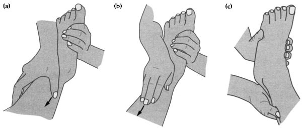
Figure 5.40 (a) Working the Spleen meridian. (b) Working the Stomach meridian between the tibia and fibula: you can also use your thumbs. (c) Working the Gall Bladder meridian and the nine terminating points of the Bladder meridian to BL-59: this is the same area as the chronic sciatic nerve.
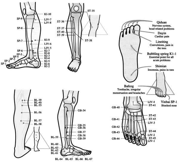
Figure 5.41 Pressure points of the lower limbs
SP-1. Regulates blood from the nose, stomach, bladder and intestines. It also calms the mind. Relieves spasms in uterus.
On the medial side of the great toe at the base of the nail.
SP-2. Promotes the digestion and helps general fatigue and agitation in children.
In the middle of the proximal phalange of the great toe.
SP-3. Strengthens the spine and helps stomach problems, hernias, lung problems. It also helps upper, middle and lower burners and helps lumbar problems.
At the distal end of the first metatarsal joint.
SP-4. This is a very complex point. It helps regulate menstrual problems and is a very useful point in all excess patterns of the stomach or spleen. Cardiac inflammation.
At the proximal end of the first metatarsal joint.
SP-5. Very good for oedema of the abdomen, constipation and any spasms of low abdomen. Constipation, haemorrhoids, spasms in the calf.
In the depression anterior to the medial malleolus.
SP-6. Vital point. DO NOT USE DURING PREGNANCY.
The next most important point next to KI-1. It is a meeting point of all three Yin points. KI-8, known as the crossing point and LIV-5. These points will stop any lower abdominal pain, whatever the cause. SP-6 is a major point for all gynaecological complaints, but it also has a strong calming action of the mind.
Four finger-breadths above the medial malleolus posterior edge of tibia.
SP-7. This point helps flatulence and abdominal swelling, also all leg problems.
Eight finger-breadths above the medial malleolus.
SP-8. Lumbar problems. Painful menstruation.
Four finger-breadths below SP-9.
SP-9 All knee problems and urinary problems and oedema of the leg. All abdominal pain.
In the depression below the head of the head of the tibia.
LIV-1. All menstrual problems. This point acts on the lower burner and helps bladder problems, constipation and also helps lumbar area.
On the lateral side of the great toe at the base of the nail bed.
LIV-2. Mainly head areas. Secondary genitals, allays insomnia and toothache. Genitourinary tract and palpitations.
In the web between the first and second toes.
LIV-3. Migraines, hypertension, mental calming point, cramp, also helps upper and lower abdominal areas.
Furthest proximal point between the first and second metatarsals.
LIV-4. Genital problems, cystitis, any paralysis.
In the depression, medial side of the crease of ankle.
LIV-5. Genitourinary problems, also colic and spasms of abdomen.
On the medial side of leg. Six finger-breadths above medial malleolus, on the midline of the tibia.
LIV-6. Knee problems and useful point for extreme pain in cystitis.
On the medial side of leg. Nine finger-breadths above medial malleolus, on the midline of the tibia.
LIV-7. Known as the Knee gate, so will help all knee problems.
Almost in line with SP-9. Slightly posterior aspect.
LIV-8. This major point will help all four limbs, all vaginal problems, discharge, pruritis, and abdominal pain.
On the medial side of leg just below SP-9 and in line with LIV-7.
BL-67. Headaches, vertigo, it is also used as a major point for malposition of the fetus in the eighth month of pregnancy. Paralysis or arthritis of foot.
On the lateral side of the little toe just below nail bed.
BL-66. Bladder problems, major point for stiff neck, headaches and uterine congestion.
On the proximal phalange of the little toe.
BL-65. Headaches, vertigo, good point for releasing necks, torticollis.
On the distal end of the fifth metatarsal joint.
BL-64. All lumbar problems, helps the bladder and calms the mind, cerebral congestion.
Just below the tuberosity of the fifth metatarsal joint.
BL-63. All pelvic problems, bladder and abdominal pain, knee problems.
On the lateral side of the foot just below the external malleolus on the proximal border of the cuboid bone.
BL-62. Major point for movement in lower back. Relaxes all tendons of legs.
In the depression immediately below the tip of the external malleolus.
BL-61. All leg problems.
On the lateral side of the calcaneum, at the point where red and white skin meet in the longitudinal line of BL-60.
BL-60. Vital point. DO NOT USE DURING PREGNANCY but may be used during labour. All backache, shoulder, neck and head problems. Sciatica, ankle and leg problems, placental retention.
On the posterior edge of the lip of the external malleolus.
BL-59. Lumbar and thigh problems, muscular and nerve problems of leg.
Four finger-breadths above the external malleolus on the tibia.
BL-58. Helps kidneys and lower leg problems.
Nine finger-breadths above the external malleolus on the tibia.
BL-57. Backache, low lumbar area. Sciatica and leg problems.
On the midline on the gastrocnemius muscle in the pointed depression.
BL-56. Lumbar problems, cramps.
Exactly six finger-breadths below popliteal crease, on the midpoint.
BL-55. As above and knee.
Roughly two finger-breadths below popliteal crease on the midpoint, above BL-56.
BL-40. Lumbago, leg problems, sciatica.
Midpoint of transverse crease of popliteal.
BL-39. Cystitis, and constipation.
Midpoint of transverse crease.
KI-1. Any acute problems, cardiac, palpitations, vertigo, sterility in females. To restore consciousness. Prolapse uterus.
On the plantar aspect of the foot between zones 2 and 3. Plantar flex the foot and the point is just below red skin.
KI-2. Cystitis, all uterus problems, orchitis, sterility, prolapse, dysmenorrhoea, incontinence of urine.
On the medial aspect of foot, just below the tuberosity of the navicular bone.
KI-3. Helps kidneys, lower back, knees, regulates uterus.
Behind the medial malleolus in the depression.
KI-4. Uterine spasms, helps and strengthens the back.
On the medial aspect of the foot behind medial malleolus and anterior to the Achilles tendon.
KI-5. All menstrual problems, helps regulate uterus.
On the medial aspect of the foot immediately below KI-3.
KI-6. Promotes function of uterus and any fluid problems, dryness in throat or eyes.
In the depression just below the tip of the medial malleolus.
KI-7. Helps all oedema. Relieves pain and strengthens lower back.
On the medial aspect of the leg, directly above KI-3.
KI-8. Regulates all menstrual problems and abdominal pain. Dissolves any masses.
Almost in line with SP-6, between the tibia and Achilles tendon.
KI-9. Extreme calming point, relaxes chest area and relieves palpitations.
Six finger-breadths above and in line with KI-3.
KI-10. Similar action to KI-8, SP-9 and LIV-8.
On the medial aspect of the popliteal crease, press when patient’s knee is flexed.
ST-45. Known as the sick mouth, all mouth problems, tonsillitis and gingivitis.
On the lateral side of the second toe at the base of nail bed.
ST-44. Lower jaw paralysis, trigeminal neuralgia, facial problems.
Just below web between second and third toes.
ST-43. Hernia, flatulence. Fevers and night sweats.
Lies on the medial side of third metatarsal in the depression between second and third metatarsals.
ST-42. Anorexia, stimulates stomach and spleen. Arthritis of the foot.
On the dorsal aspect of foot on instep where dorsalis pedis pulse is palpable.
ST-41. Constipation, headaches, rheumatism. Helps all joints especially ankles.
It is known as the dispersing stream.
In the depression midpoint of the dorsal crease of the ankle.
ST-40. Known as the abundant bridge it is next to ST-38. It aids the removal of lumps in the thyroid and uterus areas. Also a helpful point for asthma as it eliminates phlegm.
Lies on the lateral side of the tibia ten finger-breadths approx above the external malleolus. A connecting point.
ST-39. Regulates stomach and intestines, relieving any abdominal spasms. Nine finger-breadths above external malleolus on lateral side.
ST-38. Helps shoulder problems, when moving shoulder and pressing point at same time.
Six finger-breadths below the knee, lateral side of tibia.
ST-37. A good point for large intestine and very good for chronic diarrhoea. Aids lumbar and leg problems.
Five finger-breadths below the knee, lateral side of tibia.
ST-36. This is a major point to strengthen the whole body and for all abdominal problems. NOT TO BE USED ON YOUNG CHILDREN.
Four finger-breadths below the knee, lateral side of tibia.
ST-35. Stomach problems, gastritis, diarrhoea.
On lateral edge of patella.
GB-44. Alleviates headaches. Aids problems relating to eyes, ears, mouth, breast and lung areas.
On the lateral side of the fourth toe at the base of the nail bed.
GB-43. Helpful in migraine and tinnitus disorders. Aids axillary areas, arms and breast.
In the depression between the fourth and fifth metatarsals middle of proximal phalange.
GB-42. Mastitis and all axillary area. Any pain in dorsum of foot.
Distal end of fourth metatarsal on lateral edge.
GB-41. Important point helpful in relieving pain in the whole body.
In the depression between fourth and fifth metatarsals on medial edge of fifth metatarsal.
GB-40. Sciatica, leg and ankle problems. Pelvic disorders.
In the depression lateral to the tendon of the toes, in front of external malleolus.
GB-39. Weakness of limbs.
Four finger-breadths above the external malleolus.
GB-38. Lumbar problems and knee disorders, or any numbness in lower extremities.
Five finger-breadths above the external malleolus on lateral side of tibia.
GB-37. Leg problems, especially calf muscle and ankle weakness. Six finger-breadths above the external malleolus on the lateral side of tibia.
GB-36. Leg and ankle problems.
Nine finger-breadths above the external malleolus on lateral side of tibia.
GB-35. Leg problems and sciatica, particularly on fibula.
Nine finger-breadths above the external malleolus on the lateral side of fibula.
GB-34. Knee problems, chronic constipation, sciatica.
In the depression on lateral side of leg, just below the head of the fibula.
All the other reflexes are covered exactly like the right foot. Just change support hands accordingly. If there are tender areas go back and gently work them again in a different direction.
Remove the towel from the feet. Complete with relaxation techniques 20, 23 and 24 (chapter 4) as the foot should now be completely flexible. If you do not wish to massage the feet and legs finish off the session.
This is an ideal time to work the meridians as you massage. (If it is not convenient to apply cream because the client has tight trousers or does not wish to remove the relevant clothing, you can apply pressure through the clothes.) After applying a small amount of cream, use circular friction up the plantar tendon, and over the whole plantar surface of the feet. Carry on this circular movement in between all the metatarsals and on them, especially in the depression of the crease of the mid ankle. This is ST-41, a good point for any oedema. Use the knuckles to work gently around the depressions distal to the medial and lateral malleolus. Then, using the knuckles in sweeping strokes, commence up and down the spinal reflex, which is also the Spleen meridian. Also work the lateral aspect of the foot. (See here) You should now have contacted all the acupoints on the foot of the six leg meridians. You can now commence up the legs to the knees (figure 5.40).
Note. These are vital and potent acupuncture areas. Check contraindications (chapter 3) to make sure it is safe to continue working if your client is female. SP-6 and B-60 can be used only if the person is not pregnant, as they are empirical points to promote delivery during labour. If the person is menstruating it is also not advisable to work this point unless the person is aware that it creates a natural free flow of blood; sometimes this can alarm the person if they are not prepared for this.
When massaging, the client does not need to be face down. Massage also helps the lymphatic system by aiding the dispersal of any excess fluid that remains, which could cause oedema. The general blood flow and the whole circulation of the body are enhanced. Think of making three lines up the medial side of the leg on the tibia and posterior to the tibia with firm sweeping strokes. Use friction strokes over the fleshy part; this is best achieved with your hand under the client’s leg, thumbs on the medial side. Now, using one or both thumbs, or finger, work from the ankle making a pass up between the tibia (the shin bone) and the fibula (the outer bone) to the point where you cannot pass any further. At the lateral side of the tuberosity of the tibia in the depression lies ST-36; this major point strengthens the whole body. However, it is not thought suitable for use until after the teenage years are over. In the elderly or infirm it helps to normalize functions. On the lateral side of the leg, again using sweeping strokes, work right up to the knee point between the lateral tibia and the fibula, applying pressure with your thumbs as you sweep up the shin bone. The last meridian changes course from just below the calf where it then runs up right between the gastrocnemius to the popliteal fossa; this area on the calf will aid all lower back disorders, so even if you cannot finish on a massage, with the leg bent gently apply pressure through any clothing using your three fingers of either hand. For direct treatment of any acupoint use firm unmoving pressure to suit the patient’s tolerance levels for at least 30 seconds. (See figure 5.40a, b and c.)
Finish with some metatarsal flexing (see relaxation 22, chapter 4).
See relaxation 23, (chapter 4).
See relaxation 24, (chapter 4).
Metatarsal kneading and foot moulding can be used at any time together with any other relaxation technique and stretching exercises.
Details of the foot and leg meridians and acupoints are shown in figure 5.41.
1. Shake the feet; rotate the ankles both ways.
2. Wriggle and stretch your toes, fanning and separating them as much as possible (figure 5.42).
3. Extend the great toe as much as possible, pointing the toe then dorsiflexing it (figure 5.43).
4. Invert the feet (figure 5.44a), then evert the feet (figure 5.44b). (Do this exercise while sitting down.)
5. Walk on the balls of the feet; this aids the lungs and is very good for asthmatics.
6. Walk on the heels; this aids the lower back.
7. Apply pressure on the top of your foot by placing one foot on top of the other. Press the top one down and pull the bottom one up; this is very good for the spine and legs and also aids the lung area.
8. Try and pick up a pencil with your toes (figure 5.45).
9. Place a ball or other round object under your feet and roll it the length of the foot (figure 5.46); repeat two or three times.
10. For tired legs and feet, whilst in a comfortable sitting position, place a twisted towel under the arch of the foot. Holding both ends pull gently, straightening the leg and lifting (figure 5.47). This helps to stretch the Achilles tendon gently.

Figure 5.42 Wriggling and stretching toes, fanning and separating them, to help all neck- and head-related areas

Figure 5.43 Extending the great toe as much as possible, then dorsiflexing it aids neck- and head-related areas.

Figure 5.44 (a) Invert the foot inwards to help the spinal area. (b) Evert the foot outwards to help hip, knee and shoulder problems.

Figure 5.45 Pick up a pencil to aid the lungs and shoulder areas.

Figure 5.46 Place a round ball or other round object under your foot and roll it the length of your foot several times. This works all the organs at once.

Figure 5.47 For tired aching legs, place a twisted towel under the arch of your foot; pull gently whilst straightening the legs.