PARENTAL BEHAVIOR
The goal of reproduction is successful offspring. Parents want offspring who can survive the rigors of the world and produce their own descendents. Many animals produce offspring that require sustained assistance to successfully reach maturity. The particular actions that parents undertake to ensure the growth and survival of their offspring constitute parental behavior.
The extent of parental behavior in the animal kingdom occurs along a spectrum ranging from none to helicopter parents. Female salmons lay hundreds of eggs to be fertilized and then swim away. Humans are at the other end of the spectrum—investing many years and enormous resources, and perhaps hoping to be surrounded by their children until the very end.
Females in the animal kingdom do most of the parenting, although there are some exceptions. It is generally believed that males seek to fertilize as many eggs as possible, whereas females seek to successfully raise the few they sire. Parental behavior constitutes any behavior that the parent does for the offspring. For example, a pregnant dog will build a nest a day or two before giving birth. After the delivery, she will lick them clean, eat the placentas, feed them, and keep them warm (and for all this she is called a bitch). Additionally, she will aggressively defend the pups against any suspicious intruders.
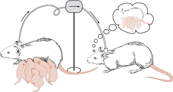
The onset of maternal behavior is remarkably precise. An inexperienced mother must immediately perform a full range of new behaviors without much room for error. How does this happen? Terkel and Rosenblatt established that there must be something in the blood that induces maternal behavior. They transfused blood from a female rat that had just delivered to a virgin rat (Figure 17.1). Within 24-hours, the virgin rat was displaying maternal behavior.
Hormones
Biologic endocrinologists have spent considerable time and energy trying to tease out the maternal molecules. Although they have gotten close, there is still no definitive concoction of hormones that will immediately trigger maternal behavior in a nulliparous (virgin) rat. The leading culprits are estrogen, progesterone, and prolactin. An important ingredient appears to be the changing levels of the hormones. In Figure 17.2, note how the progesterone is seen to drop while the estrogen and prolactin rise in a rat just before delivery.
Oxytocin also plays some important role. Traditionally, we conceptualize oxytocin as the neuropeptide released from the posterior pituitary into general circulation, which leads to uterine contractions and milk ejection (see Figure 7.4). Recent research has found receptors for oxytocin within the brain, establishing central actions for this neuropeptide. Indeed, injecting oxytocin directly into the lateral ventricles in a rat will induce maternal behavior in a hormone-primed virgin rat. More recently, it has been shown that oxytocin levels in the paraventricular nucleus (PVN) increase with maternal aggression. Likewise, infusion of synthetic oxytocin into the PVN also increases maternal aggression toward an intruder.
Further complicating this picture is the fact that hormones facilitate maternal behavior but are not required for it. Nulliparous rats will initially avoid new pups placed in their cage. However, if exposed over a series of days (1 hour each day), they will respond maternally to the pups within 5 to 6 days. Pregnant rats will show similar avoidance until after they have delivered. Then they will quickly display maternal behavior to any pup for the rest of their lives, proving that “once a mother, always a mother” (Figure 17.3).
FIGURE 17.1  When nulliparous rats are transfused with blood from a new mother, they will display maternal behavior within 24-hours. (Adapted from Nelson RJ. An Introduction to Behavioral Endocrinology. 3rd ed. Sunderland, MA: Sinauer; 2005.)
The Brain
Taken together, these studies suggest that the hormonal fluctuations late in pregnancy act on the brain to decrease fear or aversion and increase attraction toward infants. Because maternal behavior persists once it is established, it is likely that the experience permanently changes some regions in the brain. An area that has been intensely studied is the one that we discussed in Chapter 16: the preoptic area (POA) located in the anterior hypothalamus.
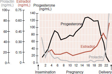
FIGURE 17.2 Blood levels of progesterone, estradiol, and prolactin in the pregnant rat. The changes that occur prior to delivery may influence maternal behavior. (Adapted from Rosenblatt JS, Siegel HI, Mayer AD. Blood levels of progesterone, estradiol and prolactin in pregnant rats. Adv Study Behav. 1979;10:225-311.)
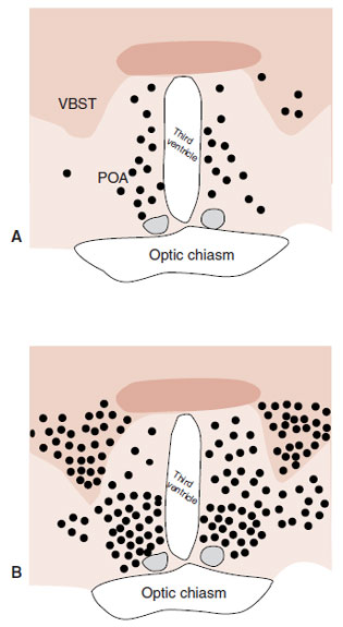
The POA is rich in estrogen, progesterone, prolactin, and oxytocin receptors, all of which increase during gestation. Lesions of the POA will disrupt maternal behavior. The POA appears to be a region that receives olfactory and somatosensory input and has projections to midbrain and brain stem nuclei. Numan and Sheehan describe an elegant experiment that demonstrates the central role of the POA with maternal behavior. Postpartum rats were exposed to either pups or candy for 2-hours. Then their brains were analyzed for the presence of the transcription factor Fos. (Fos is used as a general marker of gene expression.)
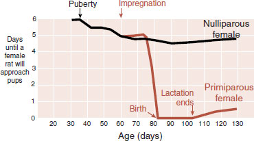
FIGURE 17.3 Female rats initially avoid pups. Several days of exposure are required for a nulliparous rat to display maternal behavior. Pregnant rats act similarly until after they deliver. (Adapted from Bridges RS. Endocrine regulation of parental behavior in rodents. In: Krasnegor NA, Bridges RS, eds. Mammalian Parenting: Biochemical, Neurobiological and Behavioral Determinants. New York, NY: Oxford University Press; 1990:93-117.)
Figure 17.4 shows the results of the study. This is a slice through the forebrain that includes the anterior hypothalamus on either side of the ventricle. (For a human comparison, see Figure 16.10.) Note the increased activation in the POA as well as other regions of the rat exhibiting maternal behavior.
Dopamine
Up to this point, we have stressed the importance of gonadal steroids and neuropeptides in the development of maternal behavior, but the neurotransmitter dopamine also appears to play an important role. We discussed in Chapter 12 the activation of the orbitofrontal cortex (OFC) and the ventral tegmental/nucleus accumbens area (see Figure 12.3) in the experience of pleasure. As might be expected, these areas are active in mothers.
One study scanned new mothers while they were looking at pictures of their own child and pictures of unfamiliar children. The mothers showed greater activation of the OFC when viewing their own child. With rats, researchers have found that mother rats will press a bar for access to pups the way they will press a bar for amphetamines or electrical stimulation. Additionally, pup exposure increases the release of dopamine at the nucleus accumbens. Alternatively, dopamine blockers will impair maternal behavior (see box). These studies give some neurobiologic explanations for the “joys of motherhood.”
Licking and Grooming
This brings us to a series of studies from Michael Meaney’s laboratory in McGill University, which may be some of the most important recent neuroscience studies for mental health professionals. These studies tie together maternal behavior with lasting effects on the offspring’s behavior, hypothalamic-pituitary-adrenal (HPA) axis, and even their DNA.
The story starts in the 1960s when researchers noted that pups “handled” once a day during the first weeks of life showed a reduced adrenocorticotropic hormone and corticosterone response to stress. Later, it was established that it was not the “handling” per se that produced this effect, but the mother’s increased licking of the pups when they were returned to the nest. The mothers were simply trying to get the human odor off their pups and this extra attention to the pups resulted in their improved response to stress when they grew up to be adults.
SCHIZOPHRENIA AND DOPAMINE BLOCKERS
Mothers who suffer from schizophrenia are known to be less involved with their children. They are generally more remote and less responsive during mother–infant play. This could be another example of the negative symptoms of the disorder. Worse yet, the problem might be exacerbated by the medications used to treat the patients.
In a recent study, Li et al. looked at the effect of injections of haloperidol, risperidone, and quetiapine on maternal behavior in rats. The antipsychotic medications inhibited maternal behaviors, such as nest building, pup licking, and pup retrieval. The figure shows the results for pup retrieval. Shortly after the injections, mothers failed to retrieve their own pups. Such studies suggest caution when treating human mothers with antipsychotic agents.
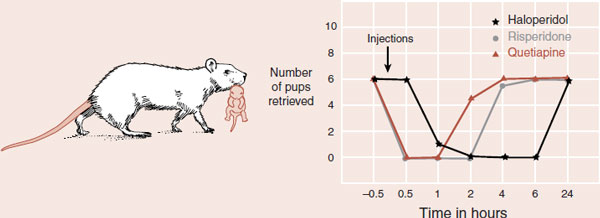
Antipsychotic medications disrupt a mother rat’s tendency to retrieve her pups. (Adapted from Nelson RJ. An Introduction to Behavioral Endocrinology. 3rd ed. Sunderland, MA: Sinauer; 2005; Li M, Davidson P, Budin R, et al. Effects of typical and atypical antipsychotic drugs on maternal behavior in postpartum female rats. Schizophr Res. 2004;70(1):69-80.)
FIGURE 17.4 Hypothalamic region of postpartum rats exposed to candy (A) or newborn pups (B). Each dot represents five cells labeled with Fos activity—a measure of gene expression. VBST, ventral bed nucleus of the terminalis; POA, preoptic area. (Adapted from Numan M, Sheehan TP. Neuroanatomical circuitry for mammalian maternal behavior. Ann N Y Acad Sci. 1997;807:101-125.)
Meaney discovered naturally occurring strains of rats that licked and groomed their pups at different rates. This particular behavior occurs when the mother rat enters the nest and gathers her pups around her for nursing. She will intermittently lick and groom the pups as they nurse. Meaney named one group the high lick and groom (high L and G) mothers and the other the low lick and groom (low L and G) mothers.
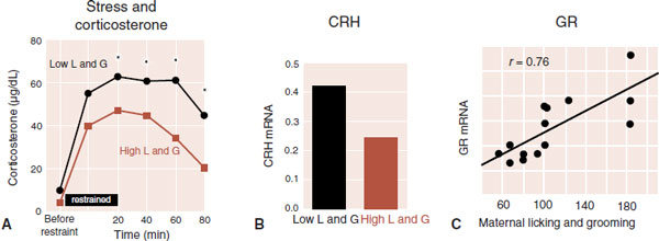
In a flurry of experiments in Meaney’s laboratory, it was established that high L and G mothers produced offspring with subtle but significantly different brains. After 20 minutes of restraint (very stressful for a rodent), the rats from high L and G mothers secrete less corticosterone (Figure 17.5A). They also produce less corticotropin-releasing hormone messenger RNA (CRH-mRNA) in the hypothalamus (Figure 17.5B). Additionally, the amount of maternal licking and grooming correlates with the number of glucocorticoid receptors (GRs) in the hippocampus (Figure 17.5C). They literally produce more GRs as a result of enhanced gene expression.
In summary, a mother’s increased attention enhances the sensitivity of the HPA axis most likely by turning on the appropriate DNA. Offspring of attentive high-licking mothers demonstrate greater feedback to the hypothalamus by way of the increased GRs, which inhibits CRH production and corticosterone release. Perhaps most significant, the pups from a high L and G mother show a greater willingness to explore novel environments as adults and demonstrate enhanced resilience under duress.
Trading Places
In a follow-up study, Meaney et al. switched some of the mothers and pups. That is, pups from high L and G mothers were raised by low L and G mothers and vice versa. The results were stunning and show how behaviors and patterns emerge from combinations of genetic predisposition and environment. Figure 17.6 shows the behavior of the adopted female rats raised by high L and G mothers once they matured. They were more inclined to explore an open area and provided greater licking and grooming to their own pups. Note how the determining factor is not the genetic makeup, but the nurturing behavior of the mother that raised them. In other words, a low L and G female will become a high L and G mother if she is raised by a high L and G mother. So the behavior can be passed from generation to generation, but it is not genetic—it is epigenetic.
Effect on the DNA
Meaney and his group have taken this line of research to the next level by searching for epigenetic mechanisms that can explain the enduring effects the mother’s behavior has on the pups. Briefly, epigenetic molecular attachments on the DNA (see Figures 6.8 and 6.9) affect gene expression, which in turn alters the proteins produced—in this case the GR.
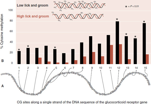
Meaney’s group identified a section of the rat DNA that encodes for hippocampal GR. Looking specifically at the promoter region of this DNA—the region where the transcription starts—they analyzed methylation of the cytosine–guanine sites (CG sites). They found a much greater frequency of methylation for the low L and G group compared with the high L and G group along the GR promoter gene (Figure 17.7). In other words, the mother’s attentive behavior reduced the methylation of the gene and allowed for greater production of the GR—which in turn made the rats more resilient. This is an excellent example of the neuroscience model (see Figure 6.13) showing how environment, gene expression, and brain development affect behavior.
FIGURE 17.5 Rats raised by a mother with a high frequency of licking and grooming behavior show a more modest corticosterone release in response to stress (A), less corticotropin-releasing hormone messenger RNA (CRH-mRNA) (B), and greater glucocorticoid receptors (GR) in the hippocampus. C. The correlation between GRs and licking and grooming by the mother. (Adapted from Liu D, Diorio J, Tannenbaum B, et al. Maternal care, hippocampal glucocorticoid receptors, and hypothalamic-pituitary-adrenal responses to stress. Science. 1997;277(5332):1659-1662.)
The researchers conducted a follow-up study with their rats that is almost unbelievable. They administered directly into the cerebral ventricles an inhibitor (trichostatin A) that results in demethylation of the DNA (Figure 17.8A). These rats displayed less methylation of their DNA and, consequently, greater numbers of GRs. Furthermore, when stressed, these animals showed a modest HPA response (Figure 17.8B); that is, their corticosterone levels became indistinguishable from those of rats raised by a high L and G mother.
The implications from these studies are profound. We can now trace the effects of a mother’s behavior down to the offspring’s DNA. Furthermore, if we can find ways to cleanse the DNA, we might be able to correct psychiatric problems, not just treat symptoms. However, it is important to note that although demethylation may be the treatment of the future, it is not without risks. Some cancers are believed to result from demethylation of growth promoting genes—genes we do not want to inadvertently turn on.
What about in humans? Meaney and his colleagues recently analyzed the human equivalent of the promoter region of the glucocorticoid gene in cells from the hippocampus in a postmortem study of human brains. They compared suicide victims with a history of childhood abuse to two control groups both without a history of abuse—a control suicide group and a control sudden death, non-suicide group. The suicide victims with a history of abuse had greater methylation of the glucocorticoid receptor gene compared with the nonabused controls. Additionally, as would be predicted, the mRNA transcribed from the GR gene was reduced in the subjects with a history of abuse. The human findings are consistent with the rat studies.
PAIR BONDING
The attraction between men and women is a special form of social attachment. In the short term, it is required for sexual reproduction; in the long run, bonded parents coordinate the rearing and protection of the offspring. Surprisingly, monogamous pair bonds are rare among mammals: approximately 5%. Monogamy is much more common among birds. The unusual bonding and rebonding that is common with humans might be best described as serial monogamy. The question for us is: what parts of the brain drive the affiliation of men and women?
Romantic Love and Dopamine
Romantic love is a universal human experience. The feeling of attraction that one person may feel for another can be intense, all-consuming, and difficult to control. A person in love feels euphoric. A spurned lover is despondent and even violent. The obsessional thinking and the willingness to “cross mountains” to be with a lover suggest the activation of the brain’s reward system (see Figure 12.6) when one is in love.
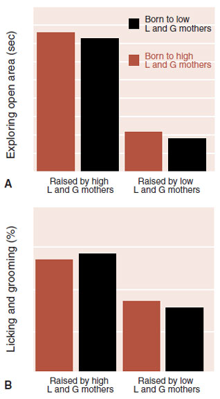
FIGURE 17.6 Female rats raised by mothers who were high lickers and groomers are less anxious in an open area (A) and are more likely to be high lickers and groomers when raising their own pups (B)—regardless of their genetic lineage. L and G, lick and groom. (Adapted from Francis D, Diorio J, Liu D, et al. Nongenomic transmission across generations of maternal behavior and stress responses in the rat. Science. 1999;286(5442):1155-1158.)
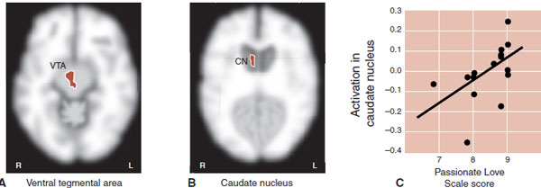
Anthropologist Helen Fisher at Rutgers University has spent her career studying the science of love. She and her colleagues scanned the brains of people who were “intensely in love.” While undergoing magnetic resonance imaging (MRI), subjects were alternatively shown pictures of their beloved or a neutral individual and the differences were measured. As expected, the images of the beloved lit up the ventral tegmental area (VTA) (Figure 17.9A). The VTA is a dopamine-rich area with projections to the nucleus accumbens. These are the subcortical regions that mediate motivation and reward.
Another area activated in the study was the caudate nucleus (CN) (Figure 17.9B). This area is also active in obsessive-compulsive disorder (see Figure 22.10). In the Fisher study, activity in the CN correlated with the total score on a test of the subject’s feelings: the Passionate Love Scale. Therefore, in simple terms, we can conceptualize love as both an addiction and an obsession.
Growth Factors
A group in Italy—a country that knows a thing or two about romance—studied growth factors in subjects who had recently fallen in love. They speculated that nerve growth factors (NGFs) might be activated when people experience romantic feelings. They drew blood from subjects who recently fell “in love” and couples in long-lasting relationships. They measured the values of four growth factors. Only one—NGF—was significantly higher in the subjects recently in love. Moreover, there was a positive correlation between the level of NGF and the subject’s score on the Passionate Love Scale.
Of particular interest, the researchers reexamined the levels of NGF a year or two later in the subjects in love. They found that the levels of NGF had dropped back to the levels seen in the control group (Figure 17.10). This is the neuroendocrine equivalent of what we all know: the honeymoon does not last. It is another example that the brain does not tolerate euphoria for too long. However, if the pleasure wanes, why do we stay in a relationship? In addition to psychological and practical answers, it may be that other neuropeptides kick in. Work with voles may shed some light on this.
Vasopressin
The vole is a rodent that looks like a plump mouse, but is related to the lemming. They are common in the grassy fields of North America. Voles are relevant to our discussion because of their diversity in forming pair bonds. For example, the prairie vole will form enduring pair bonds and mutually care for the offspring. In nature, most prairie voles that lose a mate never take on another partner. The closely related meadow (mountain, montagne) vole, on the other hand, is socially promiscuous and does not display biparental care.
In the laboratory, researchers have observed that prairie vole males prefer to spend time next to their partners: called huddling (see Figure 17.11A). The meadow vole, on the other hand, is more independent. With the proper arrangements, this behavior can be measured and quantitated (Figure 17.11B).
FIGURE 17.7 The presence of methyl groups attached to cytosine–guanine (CG) sites was assessed along the DNA of the rat glucocorticoid receptor (A). Pups from mothers who were high lickers and groomers had significantly less methylation of this section of DNA (B). (Adapted from Weaver IC, Cervoni N, Champagne FA, et al. Epigenetic programming by maternal behavior. Nat Neurosci. 2004;7(8):847-854.)
Vasopressin has emerged as a critical neuropeptide mediating the pair bond formation in male voles. Infusion of vasopressin into the male cerebral ventricles accelerated pair bond formation. Likewise, infusion of a vasopressin antagonist prevents pair bond formation. Furthermore, differences in expressions of the vasopressin receptor can be demonstrated in the two species of voles (Figure 17.11C). The prairie vole has significantly more receptors in the ventral pallidum (VP).
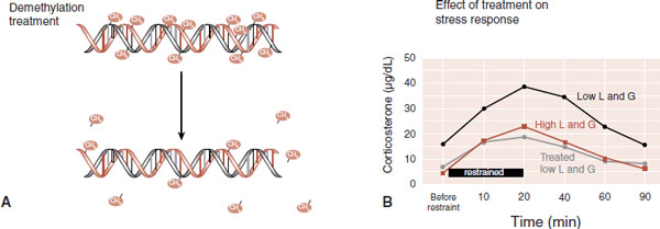
FIGURE 17.8 Trichostatin A can remove the methyl groups from DNA (A). Rats from low L and G mothers who have been treated with trichostatin A display a normal hypothalamic-pituitary-adrenal response to stress (B). (B Adapted from Weaver IC, Cervoni N, Champagne FA, et al. Epigenetic programming by maternal behavior. Nat Neurosci. 2004;7(8):847-854.)
FIGURE 17.9 Subjects intensely in love show activity in the ventral tegmental area (VTA) and caudate nucleus (CN) when looking at pictures of their lover (A and B). Activity in the CN correlated with scores on the Passionate Love Scale (C). (Adapted from Aron A, Fisher H, Mashek DJ, et al. Reward, motivation, and emotion systems associated with early-stage intense romantic love. J Neurophysiol. 2005;94(1):327-337.)
In a study that seems like something out of a science fiction novel, Young et al., through their experiments, have increased partner preference in meadow voles. They used a viral vector to transplant into the VP of meadow voles the segment of DNA that encodes for the vasopressin receptor. This resulted in increased expression of vasopressin receptors. The usually solitary meadow voles now were huddling with their partners. In essence, they changed a male from being promiscuous into being monogamous. (Won’t the social conservatives be thrilled?)
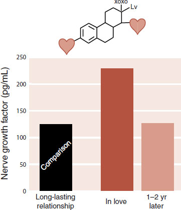
FIGURE 17.10 Nerve growth factor in subjects in long-lasting relationships, subjects actively “in love,” and these same subjects a year or two later. The hearts on the NGF molecule are the authors’ license.
With human men, we know that the endurance of pair bonding runs in families. In some families, the men partner-up for life, while in other families, the men act more like meadow voles. The role of VP in marital commitment is of great interest, but few studies have addressed the issue. One report in 2008 looked at variations of the vasopressin receptor 1a gene in over 500 pairs of Swedish twins, all of whom were married or living with a partner. Everyone answered a questionnaire on the quality of their relationship, which generated a score from 0 to 66 for each person—66 being marital bliss. Men, but not women, with one particular allele (called 334) had lower scores on the Partner Bonding Scale as measured by self-report as well as their wives’ score. The results were small but statistically significant and suggest, as with the voles, that the neuropeptide vasopressin influences pair bonding duration in humans.
Oxytocin
Oxytocin and vasopressin are remarkably similar in structure and size (Figure 17.12A). The body and brain, fortunately, recognize the subtle differences and produce distinct responses to these messengers. They are synthesized in the hypothalamus and permeate out into the body and brain by three mechanisms. The best known of these is the release of the molecules into the general circulation from the posterior pituitary (Figure 17.12B). Additionally, the molecules are released from the dendrites of the neurons and diffuse throughout the brain (dashed arrows; Figure 17.12C). Finally, small neurons project from the hypothalamus to areas such as the anterior cingulate, nucleus accumbens, and amygdala, as well as others. The essential point is that oxytocin and vasopressin are hormones as well as neurotransmitters with diverse methods of transmission and effects on physiology and behavior.
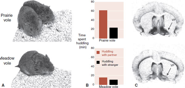
FIGURE 17.11 The male prairie vole will spend more time huddling with his partner than does the meadow vole (A and B). The difference in vasopressin receptors (dark areas in (C)) in the ventral pallidum (VP) may explain the difference in this behavior. (From Lim MM, Wang Z, Olazabal DE, et al. Enhanced partner preference in a promiscuous species by manipulating the expression of a single gene. Nature. 2004;429:754-757.)
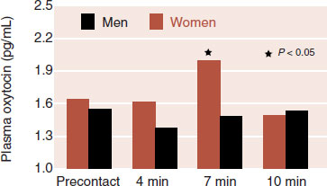
In the lay press, oxytocin is affectionately called the “cuddle hormone” since oxytocin levels are elevated with childbirth, breast-feeding, and an orgasm—all circumstances in which a woman is prone to bond with another human. In one frequently cited study, which may be the source of the “cuddle hormone” term, oxytocin significantly jumped in women after 10 minutes of “warm contact” with her spouse or partner, but did not change in men (Figure 17.13). Subsequent research using intranasal oxytocin (because it cannot be administered orally) has shown that it:
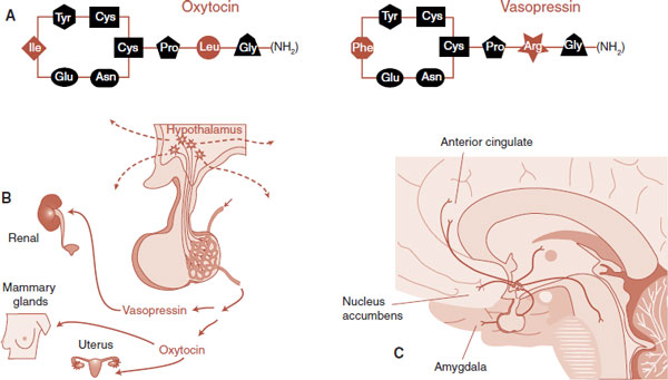
FIGURE 17.12 Oxytocin and vasopressin (A) are simple proteins consisting of nine amino acids. These neuropeptides are excreted into the pituitary circulation (B), diffuse into the brain from the dendrites of the cells (C), and are released by small neurons at several locations around the brain.
FIGURE 17.13 Oxytocin levels increase in women 7 minutes after cuddling with their partner. (Adapted from Grewen KM, Girdler SS, Amico J, et al. Effects of partner support on resting oxytocin, cortisol, norepinephrine, and blood pressure before and after warm partner contact. Psychosomatic Med. 2005;67:531-538.)
– improves positive communication between couples,
– increases trusting behavior,
– produces more positive responses of fathers toward toddlers,
– and in general enhances emotional recognition and responsiveness to others.
Brain imaging studies suggest that oxytocin may, in part, improve social interactions by dampening the amygdala/cingulate circuit—a circuit known to be activated in fearful circumstances. It might be that trust and social comfort are increased with less social anxiety.
Enduring Bonds
A simple understanding of human male–female affiliation proposes that it all starts with dopamine and the pleasure centers of the brain. We speculate that other neuroendocrine systems such as oxytocin and vasopressin may then take over to ensure enduring pair bond formation once “the thrill is gone.” While there are no data to support this as yet, twin studies suggest monogamy has biological roots. We wonder if those individuals who are prone to stay in one relationship are genetically more endowed with the neuropeptides of attachment.
DISCONNECTED
Affiliation and pair bonding are on one end of the social attachment spectrum. On the other end are those individuals who are isolated and disconnected—individuals who are aloof, distant, and fail to derive pleasure from social interactions. The Unabomber is an extreme example of this sort of person. A graduate of Harvard, with a Ph.D. in mathematics, he lived alone for 16 years in a 10 foot by 12 foot cabin in the woods of Montana without electricity or plumbing. A psychiatrist conducting a court evaluation of the Unabomber gave him a provisional diagnosis of schizophrenia.
The schizophrenic spectrum disorders include the following:
• Schizotypal personality disorder
• Schizoid personality disorder
• Delusional disorder
• Schizoaffective disorder
• Schizophrenia
These comprise a large percentage of the cases of socially disconnected individuals seen in most clinical practices. The impairment in social skills these individuals have, part of the negative signs of schizophrenia, is possibly the most troubling aspect of the illness for unaffected family members—something is missing and no treatment will bring it all back. Very little is known about the neuroanatomic deficits that contribute to the social aspect of the illnesses. However, as we will see in Chapter 23, it has been hard to define any biological deficits that explain the basic condition.
TEND-AND-BEFRIEND
Fight-or-flight has been the prevailing model to describe the mammalian response to stress. That is, stress causes a hormonal cascade that produces secretion of catecholamines and the organism either fights or retreats. Taylor has proposed that this model is male centric and does not describe how females cope in difficult times. Taylor believes females respond to stress by nurturing others and enhancing their social network, which she calls “tend-and-befriend.” Although the neuroendocrine mechanisms are the same, the behavior is different. In fact, the gender difference in affiliation is one of the most robust findings in human behavioral research.
Engh et al. provide an example from a free-ranging troop of baboons in Africa that they have been following. They have observed and recorded grooming behavior among the females. They found that females who lost a close relative experienced a significant increase in glucocorticoid levels after the death. However, they did not experience a decrease in their grooming although they had lost their close partner. Instead, other associations were established and the rate of grooming remained stable. The authors speculated that this social networking might modulate the stress response.
Kanner first described autism in 1943 at Johns Hopkins University. We now envision autism as anchoring the more extreme end of a spectrum of disorders characterized by the following:
• Severe social dysfunctions
• Early communication failure
• Presence of repetitive, rigid, and stereotypic behaviors
Asperger’s disorder and childhood disintegrative disorder are other conditions in the autism spectrum, which are believed to share common biologic foundations. These conditions may simply be a less severe form of the underlying disorder.
The social dysfunctions constitute the core deficits of the disorders. The inability to understand other people’s feelings and a failure to establish reciprocal relationships emerge early in those with autism. Robert Schultz et al. at Yale have developed techniques to study this aspect of autism. They used eye tracking technology to study spontaneous viewing patterns while watching video clips of complex social situations. They showed clips from the 1967 movie “Who’s Afraid of Virginia Woolf?” to subjects with autism and to age- and IQ-matched normal controls.
Figure 17.14 shows a drawing of one scene from the movie. In the foreground, two adults lean toward each other in a flirtatious interchange. The woman’s husband is in the background, silent, but irritated by his wife’s behavior. The eye movements from a control (brown) and an autistic subject (black) are collapsed onto this one scene. Note how the healthy control subject focuses on the eyes of the actors. Additionally, the control’s focus moves from face to face, literally outlining the charged social triangle.
The autistic subject, on the other hand, attends to less relevant aspects of the scene. This subject displays the following three findings commonly seen in subjects with autism:
• Avoidance of the eyes
• Focus on the mouth
• Preferential attention to objects rather than people
Clearly, this method of observation fails to gather the subtle social cues that are essential to understand the thoughts and feelings of other people. A quantitative assessment of the subject’s visual fixation on the eyes and mouths is shown in the graph on the right side of Figure 17.14. Note the dramatic separation between the groups with regard to eye fixation. A finding this large is seldom found in behavioral studies.
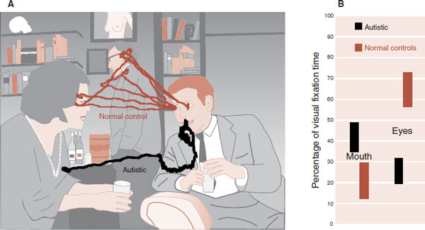
FIGURE 17.14 A drawing of a scene from “Who’s Afraid of Virginia Woolf” shows the different aspects of the movie that subjects with autism (black) and normal controls (brown) track with their eyes (A). A study comparing 15 patients with autism and 15 controls demonstrated significant differences in time spent observing the eyes and mouths from similar film clips (B). (Adapted from Klin A, Jones W, Schultz R, et al. Visual fixation patterns during viewing of naturalistic social situations as predictors of social competence in individuals with autism. Arch Gen Psychiatry. 2002;59(9):809-816; Klin A, Jones W, Schultz R, et al. Defining and quantifying the social phenotype in autism. Am J Psychiatry. 2002;159(6):895-908.)
The underlying neuroanatomic abnormalities of autism remain unknown. One consistent finding has been enlarged brain volume. In a meta-analysis, Redcay and Courchesne collected studies measuring head circumference and brain size with MRI. They calculated the percent difference from the normal for each study so that the measurements could be compared. The results are plotted by age in Figure 17.15. The authors noted that for autism the brain size is initially reduced, dramatically increases within the first year of life, but then returns to the normal range by adulthood. These findings show a period of pathologic brain growth in autism that is largely restricted to the first 2 years of life.
The phenomenon of abnormal growth in the first years of life mirrors what some parents have reported about their children, namely, that they appeared to be developing normally for the first 15 to 24 months and then showed a regression in social and/or communication skills. In a clever use of technology, Werner and Dawson had blinded observers review home video tapes of autistic children who were reported to have regressed. Indeed, they noted normal social attention and word babble at 12 months of age, but displayed significant impairment by 24 months. This was in contrast to the group of children with early-onset autism who showed impairment at 12 and 24 months of age.
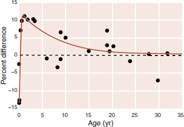
FIGURE 17.15 A meta-analysis of brain measurements including head circumference and magnetic resonance imaging scans are plotted by percent difference from norm and age. (Adapted from Redcay E, Courchesne E. When is the brain enlarged in autism? A meta-analysis of all brain size reports. Biol Psychiatry. 2005;58(1):1-9.)
Medical treatments have failed to provide any consistent benefit for the core features of autism. This is likely true because medical interventions fail to correct the synaptic dysregulation that is believed to be the underlying problem of the disorder. However, intensive behavior modification has been shown to be partially effective. One recent study with children aged 18 to 30 months found that 2 years of 20-hours a week therapist-led treatment (plus homework with the parents on the weekends) resulted in an increase in IQ of almost 18 points compared with 7 for the control group. Additionally, almost 30% in the treatment group were reclassified to a less severe form of autism after 2 years, while only 5% in the control group received a similar upgrade. These results are consistent with a basic tenet of this book—that environmental interactions (in this case B-Mod) can change the brain—possibly “rewire” some of the synaptic errors, especially when the changes are introduced early in life when the brain is more plastic and developing.
Mirror Neurons
Mirror neurons were discovered in one of those beautiful serendipitous scientific moments. Researchers in Italy placed an electrode in a neuron in a monkey’s motor cortex and noted that it was active when he grabbed an object. Much to their amazement, it also became active when the monkey watched someone else grabbing the same object. They called these neurons mirror neurons and found that they are not uncommon in the brain. Figure 17.16 shows an example of a mirror neuron. Note that the neuron is active both when a human (A) grasps the object and when the monkey (B) grasps the same object. However, the mirroring does not translate to all actions. If the object is grasped with pliers, the neuron does not become active.
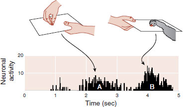
FIGURE 17.16 A neuron in the premotor cortex of a monkey is active when it observes a food morsel grasped by a human (A). The same neuron is active when the monkey grasps the morsel (B). (Adapted from Rizzolatti G, Fogassi L, Gallese V. Neurophysiological mechanisms underlying the understanding and imitation of action. Nat Rev Neurosci. 2001;2(9):661-670.)
Functional imaging studies on humans have also demonstrated mirroring. For example, a person moving a finger or observing a finger move will show similar activity in the same region of the motor cortex. Another study looked for mirroring with facial expressions. In this study, subjects were shown pictures of people displaying emotional expressions (happy, sad, angry, etc.). The subjects were instructed to either imitate the expression or just observe the picture. Figure 17.17 shows that imitating the expression and just observing it generated similar activity in the premotor region of the cortex, as measured by the functional MRI.
Additional research by this same group has identified a network of neurons connecting the frontal, parietal, and temporal lobes. Furthermore, they found that imitating emotional facial expressions not only activated this network but also activated the emotional centers of the brain, such as the insula and the amygdala. The capacity to reflect another person’s emotions may be the neuronal mechanism facilitating empathy. It may be this system that is impaired in disconnected individuals.
Dysfunction of the mirror neuronal network may underlie the lack of empathy in patients with autism. To test this hypothesis, researchers conducted similar facial imitation and observation studies with high functioning autistic children and normally developing children matched for age and IQ. Their results showed a marked decrease in activation of the mirror neuronal network in the children with autism, particularly in the frontal cortex (Figure 17.18A, B). Additional analysis showed that activity in the frontal cortex during the study correlated with the score on the social subscale of the Autistic Diagnostic Interview (Figure 17.18C).
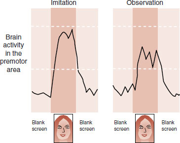
FIGURE 17.17 Activity in the facial region of the motor cortex is similar, as measured by functional magnetic resonance imaging, when subjects are imitating a facial expression or just observing it. (Adapted from Carr L, Iacoboni M, Dubeau MC, et al. Neural mechanisms of empathy in humans: a relay from neural systems for imitation to limbic areas. Proc Natl Acad Sci U S A. 2003;100(9):5497-5502.)

FIGURE 17.18 Functional MRI studies for normally developing preteens compared with high functioning age-/IQ-matched subjects with autism while imitating emotional facial expressions. The subjects with autism show less activity of the mirror neuronal network, particularly in the frontal cortex. (Adapted from Dapretto M, Davies MS, Pfeifer JH, et al. Understanding emotions in others: mirror neuron dysfunction in children with autism spectrum disorders. Nat Neurosci. 2006;9(1):28-30.)
The propensity to empathize with other’s feelings is one of the central features of human social interactions. Individuals with autism are impaired in this ability. Failing to understand the emotions in others may be a result of deficient mirror neurons in patients with autism spectrum disorders. Whether this neuronal failure is the central deficit of the disorder, a downstream effect of some other problem, or just one of a host of deficits remains to be determined.
QUESTIONS
1. Which combination of hormones is believed to trigger maternal behavior in rats just before delivery?
a. Rising progesterone and rising estradiol.
b. Rising progesterone and falling estradiol.
c. Falling progesterone and rising estradiol.
d. Falling progesterone and falling estradiol.
2. Oxytocin receptors are found in all of the following, except
a. Uterus.
b. PVN.
c. POA.
d. Optic chiasm.
3. Pups born to mothers who are high lickers and groomers show
a. Increased reluctance to explore a novel environment.
b. Increased GRs in the hippocampus.
c. Increased CRH during stress.
d. Increased corticosterone when restrained.
4. Lightly methylated DNA
a. Allows greater access to transcription factors.
b. Results in greater glucocorticoid response to stress.
c. Decreases gene expression.
d. Is induced by mothers with low licking and grooming behavior.
5. Romantic love has a strong correlation with activity in the
a. VTA.
b. CN.
c. Nucleus accumbens.
d. Amygdala.
6. All of the following are false, except
a. Vasopressin promotes pair bonding in male voles.
b. Meadow voles have more vasopressin.
c. Transplanting oxytocin receptors induces monogamous behavior in male voles.
d. Tend-and-befriend behavior modulates dips in oxytocin.
7. Eye tracking studies have shown that autistic subjects
a. Focus on the eyes.
b. Avoid looking at objects.
c. Attend to the subtle social cues.
d. Prefer to look at the lower face.
8. All of the following are true about mirror neurons, except
a. They play a role in empathy.
b. They are impaired in autism.
c. The frontosubcortical network is the most active.
d. Frontal mirror neuron inactivity correlates with social impairment in autism.
See Answers section at the end of the book.